Cherry Eye in Dogs: Causes, Treatment, and Surgery Costs
You’re playing with your dog, glancing at his face the way you always do, and you notice something that wasn’t there yesterday: a fleshy pink-red blob in the inner corner of his eye. It looks alarming. Your first instinct may be to search “red bump dog eye” at 11pm and end up reading increasingly alarming results about tumors and infections.
The good news: that red bump is almost certainly cherry eye — the common name for a prolapsed gland of the third eyelid. It is not a tumor, it is not contagious, and it is not going to cause your dog to go blind overnight. The less comforting news is that it also will not go away on its own, and the longer you wait to address it, the more likely you are to face complications.
This guide walks through everything a dog owner needs to understand: what cherry eye actually is, why it happens, which breeds are most vulnerable, what treatment looks like, realistic surgery costs in the US, and what to expect during recovery.
What Is Cherry Eye in Dogs?
The Third Eyelid and Its Role
Dogs have three eyelids — most owners only know about two. The third eyelid, formally called the nictitating membrane (from the Latin nictare, to blink), sweeps horizontally across the eye from the inner corner outward. In dogs, it is visible only at the inner corner of the eye; in some species, such as cats and birds, it is more prominent.
The nictitating membrane is not vestigial. It serves several important functions: it protects the corneal surface from debris and minor trauma, contributes to the distribution of the tear film, and — most critically — produces a significant portion of your dog’s tears. The lacrimal gland embedded within the third eyelid (the gland of the third eyelid, or nictitans gland) accounts for approximately 30–50% of the aqueous component of the tear film. That percentage is why what you do about cherry eye matters so much.
How Cherry Eye Develops
Cherry eye is, at its core, a prolapse — the nictitans gland pops out of its normal position tucked at the base of the third eyelid and becomes visible as a rounded, reddish mass at the inner corner of the eye.
The gland is normally held in place by a ligament connecting it to the periorbita (the fibrous tissue surrounding the eye socket). In dogs predisposed to cherry eye, this connective tissue attachment is congenitally weak. Under the right conditions — a sudden head movement, mild trauma, sneezing, or simply an accumulation of minor stresses — the gland prolipses forward and becomes exposed.
Once exposed, the gland is no longer properly bathed in tear fluid. It dries out, becomes irritated, and swells. The swelling makes it even harder for the gland to return to its correct position, which is why spontaneous resolution is so rare.
What Causes Cherry Eye?
Congenital Connective Tissue Weakness
Cherry eye is not caused by something the owner did or didn’t do. It is primarily a genetic structural defect in the connective tissue that anchors the nictitans gland. Dogs that develop cherry eye are born with a predisposition toward weaker anchoring ligaments — the condition is not triggered by diet, activity level, or environment.
This distinction matters practically: preventing cherry eye through lifestyle changes is not possible for at-risk breeds. The question is not whether it will happen, but whether to monitor proactively for it.
Bilateral cherry eye (both eyes affected) is common enough that veterinarians typically warn owners of an affected dog that the second eye may follow. Studies in surgical case series suggest that bilateral presentation occurs in approximately 30–40% of cases, depending on breed. In English Bulldogs, bilateral occurrence is particularly frequent.
Breeds Most at Risk
Cherry eye is strongly associated with certain breeds, particularly those with brachycephalic (short-nosed) facial anatomy and those with naturally loose facial skin. The breed predisposition reflects both the underlying connective tissue genetics and the anatomical characteristics that put additional mechanical stress on the third eyelid gland.
| Breed | Risk Level | Typical Age of Onset | Bilateral Rate |
|---|---|---|---|
| English Bulldog | Very High | 6–18 months | ~40% |
| French Bulldog | Very High | 6–18 months | ~35% |
| Cocker Spaniel | High | 6 months–2 years | ~35% |
| Beagle | High | 6 months–2 years | ~25% |
| Pug | High | 6–18 months | ~30% |
| Shih Tzu | Moderate–High | 1–3 years | ~20% |
| Bloodhound | Moderate | 6 months–3 years | ~20% |
| Lhasa Apso | Moderate | 1–3 years | ~15% |
| Boston Terrier | Moderate | 6 months–2 years | ~25% |
| Neapolitan Mastiff | Moderate | 6 months–2 years | ~20% |
Mixed-breed dogs can also develop cherry eye if they carry the relevant genetic predisposition from a parent breed. Cherry eye is less common in non-brachycephalic breeds without the characteristic loose-skinned, flat-faced anatomy, but it does occur across a wide range of dogs.
Cherry Eye Symptoms: How to Spot It
The Red Bump in the Inner Corner
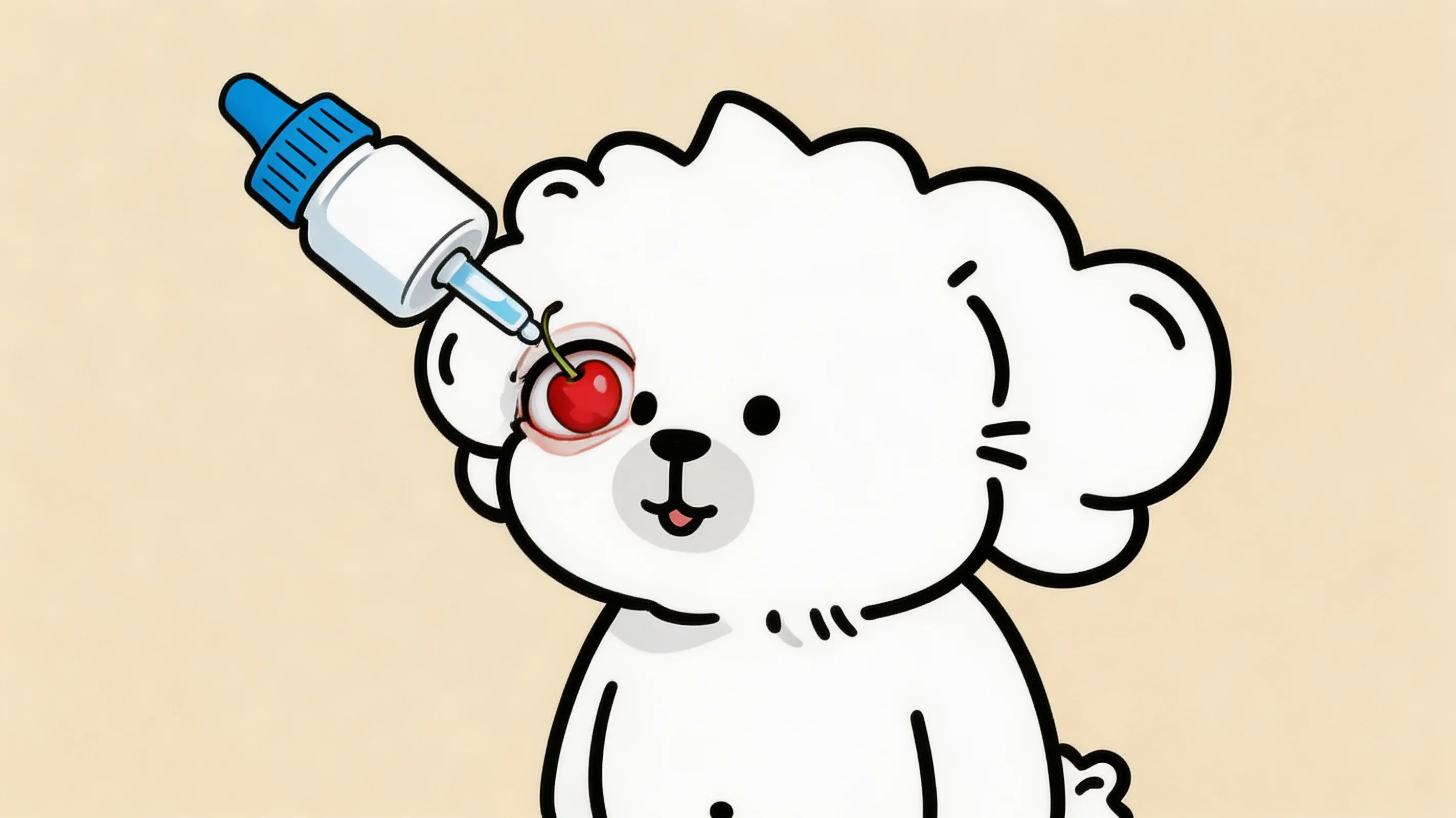
The hallmark sign of cherry eye is unmistakable once you know what to look for: a smooth, round, pinkish-red mass protruding from the inner corner (medial canthus) of one or both eyes. It typically appears suddenly — owners often report that the dog looked normal one morning and had the red mass by afternoon.
The mass can vary in size from a small pea-shaped protrusion to something closer to a small grape, depending on how long it has been prolapsed and how inflamed the gland has become. The color ranges from pale pink (if recently prolapsed and minimally inflamed) to bright red (if the gland is irritated and congested with blood).
One useful diagnostic point: unlike a foreign body or growth, cherry eye moves slightly when the dog blinks because it is attached to the mobile third eyelid. A fixed, immovable mass should be evaluated more urgently as it may represent something other than cherry eye.
Secondary Symptoms: Tearing, Redness, Pawing
Beyond the visible mass, dogs with cherry eye often develop secondary symptoms as the prolapsed gland becomes increasingly irritated:
- Increased tearing or discharge: The exposed gland produces an excess of watery or mucoid discharge. Tear staining may appear or worsen, particularly in light-coated breeds. Understanding the difference between tear staining caused by structural anatomy and tear staining caused by an underlying eye condition is important — the guide to managing tear stains in dogs covers that distinction in detail.
- Conjunctival redness: The surrounding conjunctiva (the pink membrane lining the eyelids) often becomes red and mildly swollen in response to the prolapsed gland.
- Pawing at the eye: Some dogs rub or paw at the affected eye, which can cause corneal scratches in addition to the cherry eye. An e-collar (cone) before the surgical visit may help prevent self-trauma.
- Squinting: Mild photosensitivity or surface irritation can cause the dog to squint or hold the affected eye slightly closed.
If you notice a red bump plus significant squinting, pawing, or clouding of the cornea, see your veterinarian sooner rather than later — the additional symptoms suggest surface irritation that may need immediate attention.
Is Cherry Eye Painful for Dogs?
This is one of the most common questions owners have, and it deserves a direct answer.
Cherry eye itself — the act of the gland prolapsing — is generally not acutely painful in the way a broken bone or corneal ulcer would be. Most dogs don’t cry out when it happens, and many appear unbothered in the immediate aftermath.
However, “not acutely painful” is not the same as “comfortable.” The exposed gland dries out, becomes inflamed, and causes progressive irritation and chronic low-grade discomfort. Dogs with long-standing cherry eye frequently show subtle signs of ocular discomfort: rubbing their face on surfaces, avoiding bright light, and changes in blinking behavior. If you’re uncertain whether your dog is showing pain-related behavior changes, the guide to recognizing subtle pain signs in dogs provides a practical behavioral checklist.
The discomfort tends to worsen over time as the gland becomes more inflamed, and it can increase significantly if the prolapsed gland becomes traumatized — for instance, if the dog rubs the eye repeatedly. Secondary dry eye (keratoconjunctivitis sicca), which can develop if the gland loses function, causes its own chronic discomfort and corneal irritation.
The practical takeaway: cherry eye may not be an agonizing emergency, but it is a condition causing ongoing discomfort that warrants veterinary attention — and prompt attention reduces the risk of secondary complications that cause more serious problems.
Cherry Eye Treatment Options
Conservative Management: When Eye Drops May Help
Before surgery, some veterinarians recommend a brief trial of topical anti-inflammatory eye drops (typically corticosteroid or NSAID-based ophthalmic solutions) to reduce gland inflammation and swelling. In a small subset of cases — particularly recently prolapsed glands in young dogs where the gland is not yet significantly inflamed — this can temporarily reduce the size of the mass and occasionally allows the gland to slip back into position.
The honest assessment: conservative management alone is rarely curative. Veterinary ophthalmology consensus, reflected in sources including the Merck Veterinary Manual and ACVO guidelines, is that the underlying ligament defect is structural, not inflammatory. Anti-inflammatory drops can reduce secondary swelling, but they cannot repair the weak connective tissue attachment. Most cases managed conservatively alone will recur or never fully resolve.
Eye drops may be appropriate as a short-term measure while waiting for a surgical appointment, or in elderly dogs where anesthetic risk outweighs surgical benefit — but they should not be framed as an equivalent alternative to surgery.
Will Cherry Eye Go Away on Its Own?
This is probably the most Googled question about cherry eye, so let’s answer it clearly.
No, cherry eye does not reliably resolve without treatment. Spontaneous resolution — the gland returning to its correct anatomical position without intervention and staying there — is rare. Anecdotal reports exist, but they represent exceptions, not the rule.
The reason is mechanical: the connective tissue ligament is already weakened. Once the gland has prolapsed, there is nothing to prevent it from prolapsing again. Manual reduction (massaging the gland back into place) is sometimes described as a home treatment; while some veterinarians use this as a temporary technique, it does not repair the underlying structural defect and is not recommended as a substitute for surgery.
Waiting for spontaneous resolution means prolonged exposure of the gland, increased risk of gland dysfunction, and a higher probability of secondary dry eye developing. The sooner the gland is repositioned surgically and the ligament anchored, the better the long-term outcome.
Surgical Options: Pocket Technique vs. Gland Removal
Surgery is the definitive treatment for cherry eye. Two approaches exist, and the distinction between them is clinically significant.
The pocket (envelope) technique is the gold standard and is strongly preferred by veterinary ophthalmologists and the ACVO. In this procedure, the surgeon creates a small pocket of conjunctival tissue around the prolapsed gland and sutures it closed, anchoring the gland in its correct anatomical position. The gland itself is preserved intact and continues to function.
Preserving the gland matters because of its role in tear production. Removing the nictitans gland eliminates 30–50% of the aqueous tear component permanently, substantially increasing the dog’s lifetime risk of keratoconjunctivitis sicca (KCS, or dry eye). Dry eye is a painful, chronic condition requiring lifelong daily management with lubricating drops and often prescription cyclosporine — removing the gland trades a solvable surgical problem for a permanent management burden.
Gland removal (excision) was once more commonly performed but is now considered a last resort. It is still occasionally used when the gland tissue is severely damaged, chronically inflamed beyond salvage, or has failed multiple repositioning surgeries. If a veterinarian recommends gland removal as a first-line treatment, it is reasonable to ask for a referral to a board-certified veterinary ophthalmologist for a second opinion.
Cherry Eye Surgery Cost Breakdown
Surgical cost is one of the top concerns for owners facing a cherry eye diagnosis, and it’s an area where pet health content has historically been thin. Here is a realistic breakdown based on current US market data.
What Affects the Price
The range for cherry eye surgery in the United States is broad: approximately $300 to $2,500, with most cases falling in the $500–$1,500 range. Several variables drive where a specific case lands in that range.
| Factor | Lower End | Higher End |
|---|---|---|
| Provider type | General practice vet | Board-certified veterinary ophthalmologist |
| Geographic location | Rural / lower cost-of-living area | Major metro (NYC, LA, San Francisco) |
| Number of eyes | One eye (unilateral) | Both eyes (bilateral) |
| Anesthesia complexity | Standard | High-risk breed with brachycephalic airway concerns |
| Pre-surgical diagnostics | Basic exam | Full ophthalmic workup (STT, IOP, etc.) |
A general practice veterinarian with surgical experience may perform cherry eye repair for $300–$700 per eye. A board-certified veterinary ophthalmologist (who performs dozens of these procedures annually) may charge $900–$1,800 per eye, with the higher price partly reflecting more specialized surgical technique and a lower recurrence rate.
For brachycephalic breeds — Bulldogs, Pugs, French Bulldogs — the anesthetic component of the cost may be higher because these breeds require additional precautions for airway management under general anesthesia.
Total cost for a bilateral case (both eyes) at a veterinary ophthalmologist in a major metro area can approach $2,500–$3,500 when pre-surgical diagnostics and anesthesia monitoring are included.
Does Pet Insurance Cover Cherry Eye Surgery?
The short answer: yes, in most cases — but with important conditions.
Most pet insurance policies that include illness coverage will cover cherry eye surgery, because cherry eye is classified as a disease/condition rather than a wellness service. Coverage specifics vary:
- Pre-existing condition exclusion: This is the critical caveat. If the dog was already diagnosed with cherry eye before the insurance policy was purchased — or if there is documented evidence of cherry eye symptoms in veterinary records predating the policy — most insurers will exclude it as a pre-existing condition. The timing of insurance enrollment matters enormously for conditions like cherry eye that appear in young dogs of known high-risk breeds.
- Bilateral presentation: Some policies cover both eyes as one incident; others may treat each eye as a separate claim. Review your policy language carefully if bilateral surgery is anticipated.
- Major insurers that have covered cherry eye: Trupanion, Nationwide, Healthy Paws, Lemonade Pet, and others have publicly confirmed coverage for cherry eye surgery under illness plans, subject to their standard terms. Independent review platforms like NAPHIA (North American Pet Health Insurance Association) can help compare policy terms before enrollment.
For high-risk breeds (English Bulldogs, French Bulldogs, Cocker Spaniels), enrolling in pet insurance before any symptoms appear is the most effective way to ensure cherry eye surgery is covered. If you’ve already noticed symptoms, contact your insurer before any veterinary records are created documenting the condition.
Surgery Recovery: What to Expect
Recovery Timeline Week by Week
Cherry eye surgery recovery is generally straightforward, but it does require consistent owner follow-through with eye drop schedules and activity restrictions.
Days 1–3 (Immediate post-op): The eye will be swollen and the dog may be groggy from anesthesia. Some mucoid discharge is normal. The e-collar (Elizabethan collar) is essential during this window — the dog must not rub the surgical site. Mild discomfort is expected; your vet will typically prescribe pain medication for this period. Feeding a light, easily digestible meal the evening after surgery is appropriate.
Days 4–10 (Active healing): Swelling should gradually decrease. Antibiotic eye drops (typically 3–4 times daily) and an anti-inflammatory ophthalmic ointment or drop are usually prescribed for this period. The sutures placed during surgery are typically absorbable and do not require removal. The e-collar should remain on whenever the dog is unsupervised.
Weeks 2–4 (Stabilization): Most of the visible swelling resolves. Eye drop frequency may taper down based on your veterinarian’s recheck assessment. Activity restrictions (no rough play, no swimming, no environments with dust or debris) typically continue through this period.
Week 4–6 (Follow-up and clearance): A recheck appointment at 4–6 weeks allows the surgeon to confirm appropriate healing, assess gland function, and perform a Schirmer Tear Test (STT) to verify that the repositioned gland is producing adequate tears. Most dogs receive clearance for normal activity at this point.
Post-Op Care Checklist
- Administer all prescribed eye drops on schedule (use a phone reminder if needed)
- Keep the e-collar on at all times except during supervised, calm interactions
- Check the surgical site daily for signs of infection (yellow-green discharge, significant increase in swelling, or the mass visibly re-prolapsing)
- Prevent the dog from swimming, bathing, or being exposed to dusty or dirty environments
- Limit vigorous exercise and rough play for at least two to three weeks
- Attend the scheduled follow-up recheck
Warning Signs to Watch For
Contact your veterinarian promptly if you notice:
- Yellow or green purulent (pus-like) discharge from the eye
- The red mass visibly reappearing (possible recurrence)
- Significant increase in eyelid swelling after the first 48 hours
- Persistent squinting, pawing at the eye despite the e-collar, or apparent pain
- Clouding or opacification of the cornea
Can Cherry Eye Come Back After Surgery?
Recurrence Rates and Why It Happens
Cherry eye recurrence after surgical repair is a real possibility that owners should be aware of before deciding on treatment. Published recurrence rates for the pocket technique range from approximately 5% to 20%, depending on the surgeon’s experience, the technique used, and the breed involved.
Recurrence happens when the sutures anchoring the gland to the periorbita loosen or break before adequate scar tissue forms to hold the gland in place. This is more common in breeds with particularly loose connective tissue (English Bulldogs, for instance) and when there was significant inflammation in the gland at the time of surgery.
Several factors are associated with lower recurrence rates:
- Surgery performed by a board-certified veterinary ophthalmologist
- Addressing cherry eye earlier, when gland inflammation is minimal
- Use of additional suture fixation techniques or modified approaches in high-risk breeds
What to Do If Cherry Eye Returns
If cherry eye recurs after surgery, the situation is not hopeless. A second surgery is often successful, particularly if a modified technique — additional suture anchoring, a different tissue fixation approach, or the use of a mesh material to reinforce the anchoring — is used.
Gland removal becomes a more serious consideration if multiple surgeries have failed, but even at that point, most veterinary ophthalmologists will attempt at least two repositioning procedures before recommending excision. If you are dealing with a recurrence, a referral to a board-certified ACVO ophthalmologist is particularly worthwhile — their experience with difficult cases and alternative techniques is substantially greater than that of general practice veterinarians.
What Happens If You Leave Cherry Eye Untreated?
Dry Eye (Keratoconjunctivitis Sicca)
The most serious long-term consequence of untreated cherry eye is keratoconjunctivitis sicca (KCS), commonly called dry eye. The mechanism is a direct chain from gland dysfunction to tear deficiency.
When the nictitans gland remains prolapsed and chronically inflamed, the secretory cells within the gland are progressively damaged. A gland that has been exposed and inflamed for months may lose some or all of its tear-producing capacity — even if it is eventually surgically repositioned. This is a fundamentally different situation from a gland that is promptly repositioned before significant inflammatory damage occurs.
The result is inadequate tear production. Tears are not merely lubricant — they carry oxygen and nutrients to the avascular cornea (which has no blood supply and depends entirely on the tear film) and contain antimicrobial proteins that protect the corneal surface. A dog with KCS will have chronically dry, sticky eyes with thick mucoid discharge, progressive corneal irritation, and escalating discomfort.
KCS management typically requires lifelong, twice-daily topical cyclosporine (an immunomodulatory drug that stimulates residual tear gland tissue) or tacrolimus, plus lubricating artificial tear supplementation. Dogs that develop KCS secondary to untreated cherry eye are often managed for the rest of their lives with a more intensive and expensive regimen than the surgery that could have prevented the problem.
Understanding optimal eye health nutrition for dogs — including omega-3 fatty acids that support the lipid layer of the tear film — is part of the long-term wellness picture for dogs at elevated risk of dry eye, including those with a history of cherry eye.
Corneal Damage and Vision Risk
The progression from dry eye to corneal damage is well-documented in veterinary ophthalmology. Without adequate lubrication, the corneal epithelium (the clear outer surface of the eye) begins to break down. The sequence typically runs:
- Chronic corneal dryness and irritation
- Corneal epithelial erosions (painful, shallow surface defects)
- Corneal ulceration (deeper defects with risk of infection)
- Corneal vascularization (blood vessels grow into the normally clear cornea as a healing response, reducing transparency)
- Corneal pigmentation and scarring (permanent opacification that reduces vision)
In severe, long-standing cases, corneal scarring and pigmentation can substantially reduce vision. In the worst cases — deep ulcers that perforate the cornea — vision loss can be complete and permanent in the affected eye.
This progression is not inevitable. It requires prolonged neglect of the cherry eye and subsequent dry eye. But it illustrates why treating cherry eye promptly — while the gland is still intact and functioning — produces substantially better long-term outcomes than treating it after KCS has already developed.
Cherry eye is one of those conditions that looks dramatically alarming and turns out to be medically manageable — provided it’s addressed with appropriate surgical treatment rather than left to resolve on its own. The gland of the third eyelid is a meaningful contributor to your dog’s tear production, and preserving it through the pocket technique gives your dog the best chance of normal tear film function for life.
If you’ve spotted the characteristic red mass in your dog’s eye, schedule a veterinary examination this week. Most general practice veterinarians can confirm the diagnosis and either perform the repair themselves or refer you to a veterinary ophthalmologist depending on the complexity of the case. The sooner the gland is repositioned, the better the surgical outcome and the lower the long-term risk of dry eye.
FAQ
Can I push cherry eye back in myself?
Is cherry eye an emergency?
Can cherry eye happen in one eye and then the other?
How long does cherry eye surgery take?
Can puppies get cherry eye?
Related Articles
7 Silent Signs of Cat Arthritis Most Owners Miss
Cat arthritis symptoms are easy to miss because cats hide pain instinctively. Learn the 7 behavioral signs, how vets diagnose feline joint disease, and what you can do at home.
Dog Skin Allergies: Types, Symptoms, and Management Guide
Dog skin allergies: 3 types, symptom patterns by location, 8–12 week elimination diet protocol, and treatment options including JAK inhibitors.
Dog Arthritis: Symptoms and Management
Recognize early signs of arthritis in dogs and learn effective management strategies for pain relief and improved mobility.
Dog Cruciate Ligament Tear: 7 Things Every Owner Must Know
Dog cruciate ligament tear guide: recognize symptoms, compare TPLO vs TTA surgery, follow a phased recovery timeline, and protect the opposite knee.
Dog Ear Infections: Causes, Symptoms, Treatment & Prevention
Dog ear infection causes, symptoms, and treatment: bacterial vs yeast differences, high-risk breeds, step-by-step ear cleaning, and when to see a vet.
Is Your Dog Showing Signs of IVDD? Symptoms, Stages, and What to Do
Learn to recognize IVDD in dogs symptoms by grade and spinal region, understand treatment options from conservative care to surgery, and manage recovery at home.
Dog Hip Dysplasia: 6 Essential Facts Every Owner Should Know
Learn the key facts about dog hip dysplasia — from at-risk breeds and early symptoms to PennHIP vs OFA diagnosis and home care exercises.
Why Is My Dog Limping? A Cause-by-Cause Diagnosis Guide
Dog limping on front or back leg? Learn the most common causes by leg and age, a home observation checklist, and when the limp needs emergency care.
If Your Dog's Hind Legs Are Getting Thinner, It Could Be Sarcopenia
Dog muscle atrophy in hind legs is an early warning sign of sarcopenia. Learn the stages, causes, BCS/MCS self-assessment, and evidence-based recovery strategies.
The Hidden Link Between Your Dog's Weight and Joint Health
Extra weight does far more than strain your dog's joints — it actively inflames them. Learn the dual-pathway science and a practical roadmap to protect joint health.
Dog Periodontal Disease: Symptoms, Stages, and What Every Owner Should Know
Learn the 4 stages of dog periodontal disease symptoms, warning signs, treatment options, and daily home care steps to protect your dog's teeth and overall health.
Puppy Growth Plate Care: Safe Exercise and Breed Timelines
Learn how to protect puppy growth plates with breed-specific closure timelines, age-based exercise limits, and nutrition guidelines. Evidence-based guide.
Complete Guide to Patellar Luxation in Dogs
Learn about the causes, symptoms, grades, treatment options, and prevention of patellar luxation in dogs.
Cat FLUTD: Symptoms, Causes, Treatment, and Prevention Guide
Comprehensive guide to cat FLUTD — covering feline lower urinary tract disease symptoms, causes, treatment options, cost expectations, and home prevention strategies.
A Veterinary Guide to Cat Dental Health: From Home Care to Professional Treatment
Protect your cat's dental health with vet-backed guidance on periodontal disease, FORL, stomatitis, a 4-week toothbrushing protocol, and cleaning criteria.
Kennel Cough in Dogs: Symptoms, Treatment, and Prevention Explained
Recognize kennel cough symptoms, distinguish it from tracheal collapse and heart disease, and learn which vaccines offer the best protection for your dog.
Why Is My Dog Reverse Sneezing? Causes, Remedies, and When to See a Vet
Learn what causes reverse sneezing in dogs, how to distinguish it from tracheal collapse or kennel cough, and when the symptom warrants a vet visit.
Cat Vomiting: 7 Common Causes, Color Guide, and When to See a Vet
Understand cat vomiting causes — from hairballs to serious illness. Includes a full vomit color chart with urgency ratings and a step-by-step home care guide.
Cat Constipation: More Water Won't Always Fix It — Causes, Remedies, and When to See a Vet
Cat constipation causes and remedies: 7 causes, 3-stage severity guide, home care with belly massage and pumpkin, Miralax dosing, and vet visit thresholds.
Why Does My Cat Have Diarrhea? Causes, Stool Color Guide, and When to Act
Discover the 7 most common cat diarrhea causes, decode stool colors with our risk chart, learn safe home remedies, and know exactly when to call your vet.
Cat Diabetes Symptoms, Treatment, and Remission: A Complete Guide
Recognize cat diabetes symptoms early, understand insulin and SGLT2 treatment, get a realistic cost breakdown, and learn what remission really means.
Dog Diarrhea Causes: Color Guide and Day-by-Day Home Care
Dog diarrhea causes explained by stool color, with a day-by-day home care protocol, vet warning signs, and weight-based pumpkin dosages.
5 Causes of Bad Breath in Dogs and What Each Smell Means
Dog bad breath causes span dental disease to kidney failure. Decode 5 root causes by smell type, with a home check protocol and vet-backed remedies.