7 Types of Lumps on Dogs: How to Tell Benign from Malignant and What to Do Next

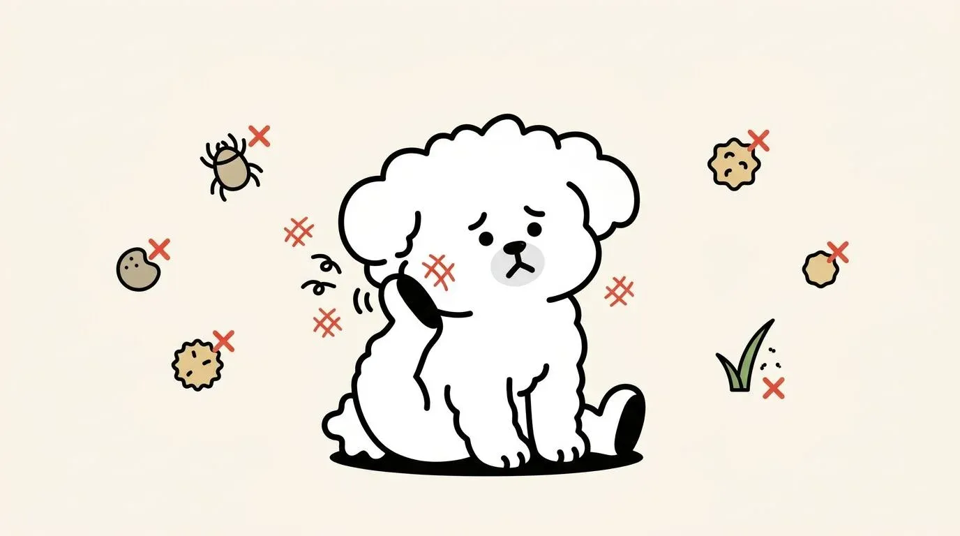
Finding a lump on your dog during a routine belly rub is one of the most unsettling experiences a pet owner can have. The instinct to panic is understandable — but most lumps on dogs are not cancer.
According to the Merck Veterinary Manual, benign growths account for the majority of skin masses seen in veterinary practice, with lipomas alone making up a significant portion. That said, distinguishing the harmless from the dangerous is not something you can do reliably at home. This guide walks through the 7 most clinically significant types of lumps on dogs, the checkpoints that matter most, what diagnostic tests cost, and how to move forward confidently.
Why Dogs Get Lumps, Bumps, and Skin Growths
Canine skin is a complex, active organ — and over a dog’s lifetime it accumulates cellular errors, hormonal changes, viral exposures, and chronic friction. Any of these can produce an abnormal growth.
What Is the Difference Between a Mass and a Tumor?
These terms are often used interchangeably, but they carry different clinical weight. A mass is a nonspecific term for any palpable lump, regardless of its nature. A tumor (from the Latin for “swelling”) technically means any abnormal tissue proliferation, but in clinical use it usually implies a growth driven by uncontrolled cell division — which may be benign or malignant.
Benign tumors grow locally, do not invade surrounding tissue, and do not spread (metastasize) to other organs. Malignant tumors (cancer) do the opposite: they invade locally, may travel through lymph nodes or the bloodstream to distant organs, and are capable of killing the host.
The distinction cannot be made by feel, appearance, or owner observation alone. It requires cellular examination by a veterinary pathologist.
Breeds and Ages Most Prone to Skin Lumps
Age and genetics both influence which growths a dog is likely to develop. Cornell University’s College of Veterinary Medicine notes that middle-aged to senior dogs (6 years and older) carry the greatest overall risk for skin tumors.
| Breed | Elevated Risk | Notes |
|---|---|---|
| Labrador Retriever | Lipoma | Very high frequency; often multiple |
| Boxer | Mast cell tumor | Among highest MCT rates of any breed |
| Golden Retriever | Mast cell tumor + lipoma | ~60% lifetime cancer incidence |
| Pug | Histiocytoma | Common in younger Pugs |
| Cocker Spaniel | Sebaceous cyst, lipoma | Glandular predisposition |
| Scottish Terrier | Bladder + skin tumors | Generally high cancer risk |
| Bernese Mountain Dog | Histiocytic sarcoma | Breed-specific aggressive malignancy |
Young dogs under 3 years most commonly develop histiocytomas — button-like, rapidly appearing benign growths that frequently resolve on their own within 2-3 months. Senior dogs, particularly in high-risk breeds, are more likely to present with lipomas, sebaceous cysts, and occasionally malignant skin tumors.
7 Common Types of Lumps on Dogs
Most skin lumps fall into a manageable set of categories. Rather than cataloging every possible growth (PetMD lists 22 types), this guide focuses on the 7 you are most likely to encounter and the ones with the highest clinical stakes.
Lipoma: Soft, Fatty Lumps Under the Skin
What it is: A benign tumor of fat cells (adipocytes) that accumulates in the subcutaneous (under-skin) layer. Lipomas are the most common benign soft-tissue tumors in dogs.
Appearance and feel: Soft, smooth, dome-shaped, and freely movable when you press it. The skin over the lump slides easily. Typically 1-5 cm in diameter, though some grow considerably larger.
Common locations: Chest wall, ribcage, upper legs, abdomen.
Risk level: Low. Lipomas rarely become malignant. The exception is the infiltrative lipoma — a variant that grows into surrounding muscle — which is harder to remove completely and can recur.
Key concern: A lipoma growing in a confined space (between muscle layers, near a joint) can cause functional problems despite being benign.
Sebaceous Cyst: Blocked Oil Gland Bumps
What it is: A cyst formed when a sebaceous (oil) gland or hair follicle becomes blocked, trapping keratin and sebum (oily secretion) beneath the skin.
Appearance and feel: Round, firm to soft, with a clear boundary. Often has a dark central pore (the blocked follicle opening). May contain a thick, white, cottage cheese-like material. Usually 0.5-2 cm.
Common locations: Back, neck, limbs — anywhere hair follicles are dense.
Risk level: Low. Sebaceous cysts are benign but prone to rupture and secondary infection. A ruptured cyst becomes inflamed and painful and requires veterinary treatment. Breeds with oily skin (Cocker Spaniels, Basset Hounds) are predisposed.
Papilloma (Warts): Viral Skin Growths
What it is: Benign viral growths caused by canine papillomavirus (CPV). They appear most commonly in puppies and immunocompromised adult dogs.
Appearance and feel: Cauliflower-like or frond-shaped (multiple finger-like projections), pink-gray, and clustered. Most common around the mouth, lips, and eyelids; occasional appearance on the feet or genitals.
Risk level: Low. Most warts resolve spontaneously within 1-5 months as the immune system clears the virus. Persistent warts in immunocompromised dogs may require removal.
Contagion note: Canine papillomavirus is not transmissible to humans, but can spread between dogs through direct contact.
Skin Tags: Small, Harmless Projections
What it is: Small, soft, pedunculated (stalk-attached) growths of fibrous skin tissue. The medical term is acrochordon or fibrovascular papilloma.
Appearance and feel: Hanging, floppy, skin-colored or slightly pigmented, 1-10 mm. Soft and easily compressed.
Common locations: Areas of friction — armpits, groin, neck collar line, between toes.
Risk level: Very low. Skin tags are entirely benign. They may become irritated if located in high-friction areas or if the dog chews at them. Removal is elective but straightforward.
Mast Cell Tumor: The Most Common Malignant Skin Tumor
What it is: A malignant tumor arising from mast cells — immune cells found throughout the skin that normally play a role in allergic response. Mast cell tumors (MCTs) are the most frequently diagnosed malignant skin tumor in dogs, accounting for 16-21% of all canine skin tumors according to veterinary oncology literature.
Appearance and feel: Highly variable — which is what makes MCTs dangerous. They can appear as small, flat, soft, skin-colored lesions or as raised, ulcerated, firm nodules. An MCT may shrink temporarily (due to mast cell degranulation), then enlarge again — this fluctuation is a significant warning sign.
Common locations: Anywhere on the body, but trunk, perineum, and limbs are most common.
Risk level: High. MCTs are graded on a histological scale (I, II, III or low/high grade). High-grade MCTs are aggressive and metastasize readily to lymph nodes, spleen, and liver.
Warning signs: A lesion that swells dramatically after touching or manipulation (Darier’s sign in dogs) may be releasing histamine from mast cells — a tell-tale indication to stop handling and see a vet promptly.
Melanoma: Dark-Pigmented Growths
What it is: A tumor of melanocytes (pigment-producing cells). In dogs, melanoma behavior varies dramatically by location: oral and nail-bed melanomas are highly malignant, while most cutaneous (skin surface) melanomas are benign.
Appearance and feel: Flat or slightly raised, darkly pigmented (brown, gray, black). May be hairless over the growth.
Common locations: Skin, mouth, nail bed, eye.
Risk level: Location-dependent. Skin melanomas are usually benign; oral and subungual (nail-bed) melanomas are among the most malignant tumors in dogs, with high rates of lymph node and lung metastasis.
Squamous Cell Carcinoma: Surface Skin Cancer
What it is: A malignant tumor arising from squamous epithelial cells — the outer layer of the skin. Chronic UV exposure is a known risk factor, making lightly pigmented, sparsely coated skin areas most vulnerable.
Appearance and feel: Irregular, wartlike, or ulcerated surface. Often crusty or bleeds on contact. Tends to be firm and fixed.
Common locations: Abdomen (sparse hair), ears, nose, nail beds, toe tips.
Risk level: High. SCC invades locally and can metastasize to lymph nodes. Early surgical removal offers the best prognosis. Delayed treatment significantly worsens outcomes.
At-a-Glance Comparison Table
| Type | Benign/Malignant | Texture | Mobility | Most Common Location |
|---|---|---|---|---|
| Lipoma | Benign | Soft, smooth | Freely movable | Chest, ribs, abdomen |
| Sebaceous cyst | Benign | Firm to soft | Movable | Back, neck |
| Papilloma (wart) | Benign | Rough, cauliflower | Non-mobile | Mouth, lips, eyelids |
| Skin tag | Benign | Soft, floppy | Pedunculated | Friction areas |
| Mast cell tumor | Malignant | Variable | Variable | Trunk, perineum |
| Melanoma | Location-dependent | Flat to raised | Variable | Mouth, nail bed, skin |
| Squamous cell carcinoma | Malignant | Firm, irregular | Fixed | Abdomen, ears, nose |
Benign vs Malignant: 5 Checkpoints Every Owner Should Know
No home observation can replace veterinary diagnosis — but knowing what to look for helps you communicate accurately with your vet and decide how urgently to seek care. Check your dog monthly using these five criteria.
Growth Rate
Benign growths tend to grow slowly over months to years. A lump that doubles in size within 2-4 weeks is a significant red flag. Conversely, histiocytomas appear rapidly (within days) but are benign — the exception that reinforces why rapid growth alone is not diagnostic.
What to do: Photograph the lump with a ruler for scale and date each photo. This gives your veterinarian objective data.
Shape and Color Uniformity
Benign growths are generally smooth-bordered and uniformly colored. Malignant growths may have:
- Irregular, poorly defined borders
- Mixed coloration (darker or lighter patches within the same lesion)
- Satellite lesions appearing nearby
The classic skin cancer acronym ABCDE (Asymmetry, Border irregularity, Color variation, Diameter >1 cm, Evolution/change) — originally developed for human melanoma — provides a useful general framework for dogs as well.
Texture and Mobility
A freely movable lump is more likely to be benign (lipoma, cyst). A lump that feels fixed to underlying muscle or bone — meaning you cannot slide it freely under the skin — is more concerning for malignancy.
Texture alone is unreliable: mast cell tumors can feel soft and movable; some cysts feel quite firm. Do not attempt to conclude “benign” based on softness.
Surrounding Skin Changes
Examine the skin around the lump, not just the lump itself:
- Redness and warmth: Can indicate inflammation or infection, but also MCT-related histamine release
- Ulceration or bleeding: Any open sore on or around a growth warrants same-week veterinary assessment
- Hair loss over the lump: Common with both benign cysts and malignant tumors — not diagnostic on its own
- Swelling spreading beyond the lump border: Suggests invasive growth or secondary infection
Your Dog’s Behavior
Changes in your dog’s behavior may be the earliest sign that a lump is causing problems beyond the skin. Watch for:
- Repeated licking, scratching, or chewing at the lump site
- Guarding the area when touched
- Changes in gait or posture if the lump is near a joint or limb
- General signs of illness: lethargy, reduced appetite, weight loss
Behavioral changes associated with pain warrant early veterinary attention. If you’re unsure whether your dog is in discomfort, the guide on dog pain behavior signs offers a detailed breakdown of subtle and overt pain indicators.
Veterinary Diagnosis: What to Expect and How Much It Costs
Discovering a lump is the trigger for a vet visit — not a diagnosis. Here is what the workup typically involves and what it costs in the US.
Fine Needle Aspiration (FNA): The First Step
What it is: A thin needle is inserted into the lump to withdraw a small sample of cells, which are then examined under a microscope. The procedure takes minutes and usually requires no sedation.
Cost: $100-$300 (includes pathologist fee if sample is sent to an external lab)
Accuracy: FNA is highly accurate for lipomas, cysts, and round cell tumors (including MCTs). It is less reliable for sarcomas and some solid tumors where cells do not exfoliate well. A non-diagnostic FNA does not mean the lump is safe — it means a biopsy is needed.
What your vet will do first: Many vets perform an in-house cytology (examining the sample themselves) before sending it to a pathologist. This can provide a preliminary impression within the same visit.
Biopsy: Definitive Diagnosis
What it is: A surgical sample of the lump — either partial (incisional biopsy) or complete removal (excisional biopsy) — is sent to a veterinary pathologist for histopathological examination.
Cost: $300-$800 (procedure + pathology lab fee)
When it is needed: When FNA is non-diagnostic, when the growth is being surgically removed anyway, or when the clinical picture suggests a diagnosis that requires histopathology (e.g., MCT grading, SCC staging).
Turnaround: Pathology results typically return in 3-7 business days.
Imaging: Checking for Spread
If a malignant diagnosis is confirmed or strongly suspected, imaging is used to assess whether the cancer has spread.
| Test | Purpose | Estimated Cost |
|---|---|---|
| Chest X-ray | Check for lung metastasis | $200-$400 |
| Abdominal ultrasound | Evaluate spleen, liver, lymph nodes | $300-$500 |
| CT scan | Detailed staging, surgical planning | $1,500-$3,000 |
| Fine needle of lymph node | Assess regional spread | $100-$200 |
This is also a useful time to schedule a comprehensive health checkup, as senior dogs presenting with a new mass benefit from baseline bloodwork and urinalysis to assess overall health before any anesthesia.
Treatment Options and Surgery Costs
Treatment depends on the diagnosis, grade, and location of the growth — and on the dog’s overall health.
When Monitoring Is Enough
Not every lump requires immediate treatment. Watch-and-wait is appropriate when:
- FNA confirms a benign lipoma that is small (<3 cm), not interfering with movement, and not in a sensitive location
- A histiocytoma is confirmed in a young dog (most resolve within 3 months without treatment)
- A skin tag is present in a low-friction area and the dog is not bothering it
Monitoring means monthly measurement and photography, plus vet reassessment every 3-6 months. Any change in size, texture, or your dog’s behavior overrides the monitoring plan.
Surgical Removal
Surgery is the primary treatment for most localized skin masses — both benign and malignant.
| Scenario | Estimated Cost |
|---|---|
| Benign lipoma removal (small, accessible) | $300-$800 |
| Benign lipoma removal (large or deep) | $800-$1,500 |
| Malignant tumor excision with wide margins | $1,000-$3,000 |
| Complex surgery (limb, face, digit) | $2,000-$5,000+ |
Surgical margin: For malignant tumors, surgeons aim for a clear margin — normal tissue on all sides of the removed tumor. MCTs require a minimum 2-3 cm lateral margin and one fascial plane deep. Incomplete excision significantly increases recurrence risk and may require a second surgery.
For infiltrative lipomas that grow into muscle tissue, wide surgical excision is more challenging, and recurrence rates are higher. Your vet may refer you to a veterinary surgeon for these cases.
For post-operative recovery guidance, the dog post-surgery recovery care guide covers wound care, activity restriction, and the suture timeline in detail.
Treatment costs add up quickly — and pet insurance can cover a significant portion of diagnosis and surgical expenses. Most plans require enrollment before a diagnosis is made, which is another reason to consider coverage while your dog is young and healthy.
Chemotherapy and Radiation
For malignant tumors that cannot be surgically removed, or where there is evidence of spread, systemic or local therapies may be recommended.
| Therapy | Indication | Estimated Cost |
|---|---|---|
| Chemotherapy (oral or IV) | MCT (high-grade), lymphoma, SCC with spread | $3,000-$10,000+ (full protocol) |
| Radiation therapy | Incomplete excision margins, MCT, SCC | $6,000-$15,000 (full course) |
| Targeted therapy (Palladia/toceranib) | High-grade MCT | $1,500-$4,000/year |
A board-certified veterinary oncologist manages these therapies. Your primary vet can provide a referral. While the costs are significant, many owners find that staged payment plans through veterinary oncology practices and partial insurance reimbursement make treatment feasible.
Recovery After Lump Removal Surgery
Regardless of whether the removed lump was benign or malignant, the immediate post-operative period requires attentive home care.
Week 1 Post-Surgery Care
The first 7 days are the most critical for wound healing and complication prevention.
E-collar (cone of shame): Non-negotiable. Dogs will lick surgical sites given any opportunity, introducing bacteria and disrupting sutures. Keep the e-collar on at all times, including during sleep. Inflatable collars may be used for small dogs where a rigid cone interferes with eating, but confirm with your vet.
Incision monitoring: Check the incision twice daily. Normal post-operative appearance includes mild swelling and bruising around the edges for the first 2-3 days. Contact your vet immediately if you see:
- Active bleeding or discharge beyond the first 24 hours
- Green, yellow, or foul-smelling discharge
- Separation of wound edges (dehiscence)
- Increasing redness or warmth extending beyond the incision
Activity restriction: Strict rest means leash walks only (2-3 minutes to eliminate), no running, no jumping, no rough play. Most surgical sites require 2 weeks of restricted activity. Swimming and bathing are prohibited until sutures are removed and the wound is sealed.
Medications: Administer prescribed pain medication and antibiotics as directed — even if your dog appears comfortable. Do not discontinue antibiotics early.
Monitoring for Recurrence
Once the immediate post-operative period passes, long-term monitoring begins.
- First follow-up: 10-14 days post-surgery for suture removal and incision check
- Pathology results: If not yet received, your vet will review the histopathology report at this visit and discuss grade, margins, and next steps
- For malignant tumors: Recurrence monitoring schedule varies by tumor type; MCTs are typically re-evaluated at 1, 3, and 6 months post-surgery
- Photograph the scar site monthly so you can compare if any new growth appears at the same location
How to Prevent Skin Lumps in Dogs
Not all lumps are preventable — genetics and aging play powerful roles. But there are evidence-based steps that reduce the risk of certain growths.
Weight Management and Diet
Obesity is a documented risk factor for lipoma development in dogs. Excess adipose tissue creates more opportunities for abnormal fat cell proliferation. Dogs maintained at a lean body condition score (4-5 out of 9 on the Purina body condition scale) develop fewer lipomas than overweight counterparts in clinical observations.
Practical steps:
- Feed measured portions rather than free-feeding
- Use a body condition scoring chart quarterly to assess your dog’s weight visually
- Choose a diet appropriate for your dog’s life stage and activity level
Skin health supplements — particularly omega-3 fatty acids from fish oil — support overall skin barrier function and may reduce sebaceous gland dysfunction that contributes to cyst formation. This is an area of active discussion in integrative veterinary medicine, though direct evidence for cyst prevention is limited.
For dogs with existing skin conditions or allergies, working with your vet to control chronic inflammation reduces the cumulative cellular stress that increases tumor risk over time.
Monthly Skin Check Routine
The single most effective prevention tool is systematic early detection. Run through this routine once a month:
- Head and ears: Feel along the skull, behind the ears, and along the jaw
- Neck and shoulders: Part the fur and check skin surface, not just palpate
- Chest and armpits: Lift each front leg and check the axillary (armpit) region
- Abdomen: Examine lightly pigmented skin particularly closely — SCC risk area
- Back and flanks: Run both hands from neck to tail base with gentle pressure
- Hindquarters: Check the perineal area (around the tail base) — MCT hotspot
- Legs and paws: Check between digits, nail beds (melanoma site), and the pads
- Record: Date and note any new findings. Photograph anything new
Any new finding that persists more than 2 weeks should be evaluated by a veterinarian.
Early Spaying and Mammary Tumor Prevention
Mammary (breast) tumors are among the most common cancers in intact female dogs. According to veterinary oncology research, spaying a female dog before her first heat cycle reduces her lifetime risk of mammary tumor development by more than 99%. Spaying before the second heat still reduces risk by approximately 92%, but the protective effect diminishes with each subsequent cycle.
If you have an intact female dog, early spaying is the most powerful single intervention available for cancer prevention. Note that this specifically prevents mammary tumors — it does not significantly affect risk for MCT, lipoma, or other skin growths.
Sun protection is an underutilized preventive strategy for dogs at risk of squamous cell carcinoma — particularly white-coated dogs with sparse belly fur who spend significant time outdoors. Pet-safe sunscreen applied to the belly and ear tips before prolonged sun exposure is a practical step for these dogs.
Lumps on dogs are common, and most are benign — but the only way to know for certain is through veterinary diagnosis. The home assessment checkpoints in this guide are a starting point for monitoring, not a substitute for professional evaluation. Monthly skin checks, maintaining a healthy weight, breed-aware risk awareness, and prompt veterinary attention for any growth that changes or concerns you are the foundations of responsible care.
References
- 1. Mast Cell Tumors in Dogs - Merck Veterinary Manual
- 2. Lumps, Bumps, Cysts & Growths on Dogs - Cornell University College of Veterinary Medicine
- 3. Skin Tumors - American Veterinary Medical Association (AVMA)
- 4. Canine Cutaneous Mast Cell Tumors: Diagnosis and Treatment - Veterinary Clinics of North America
- 5. Lipomas in Dogs - PetMD
FAQ
Are cancerous lumps on dogs hard or soft?
When should I worry about a lump on my dog?
Can I pop a lump on my dog?
How can I shrink a lipoma on my dog naturally?
Are dog warts contagious to humans?
What causes sudden lumps under a dog's skin?
Related Articles
Dog Skin Allergies: Types, Symptoms, and Management Guide
Dog skin allergies: 3 types, symptom patterns by location, 8–12 week elimination diet protocol, and treatment options including JAK inhibitors.

[2026] Dog Wellness Exam Guide: Schedule, Costs, Tests & How to Prepare
Everything dog owners need to know about wellness exams — AAHA life-stage schedule, what's included, real cost ranges, blood test interpretation, and how to prepare.

Dog Post-Surgery Care: A 2-Week Recovery Plan That Works
Dog post surgery care guide: Day 0 to Day 14 timeline, incision monitoring, medication tips, cone alternatives, and emergency warning signs.