Dog Cruciate Ligament Tear: 7 Things Every Owner Must Know
Your dog was running normally one moment and suddenly holding up a back leg the next. Or maybe the limp has been subtle for weeks, coming and going, and you have been hoping it would just resolve on its own.
A dog cruciate ligament tear is the single most common orthopedic injury seen in veterinary practice — more common than hip dysplasia, more common than patellar luxation, and far more likely to require surgery. It can happen to any breed, any age, and any size of dog, though certain factors make some dogs far more vulnerable than others.
This guide covers everything you need to make an informed decision: what the injury actually is, how to recognize it, why surgery is usually necessary for larger dogs, how to compare your surgical options, and what a realistic recovery looks like week by week.
What Is a Cruciate Ligament Tear in Dogs?
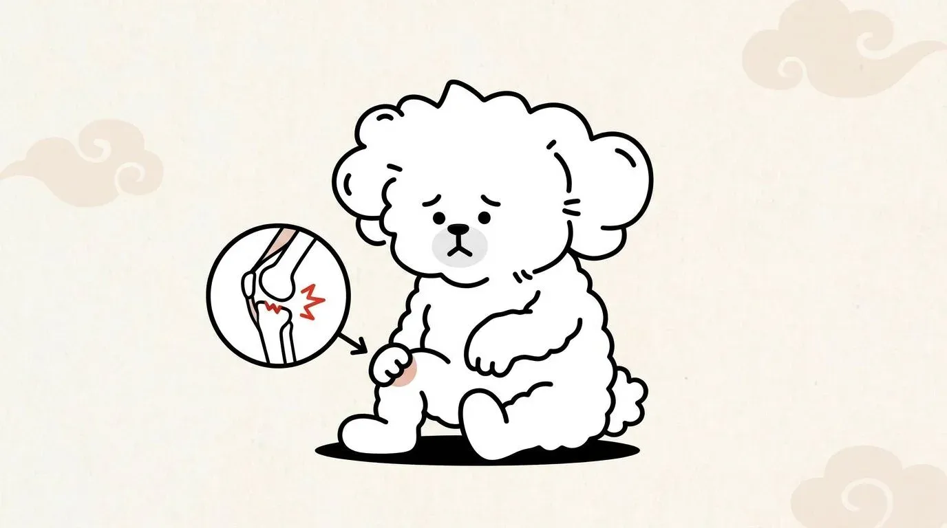
The knee joint in dogs — called the stifle — is held together by two crossing ligaments inside the joint capsule. The one that almost always tears is the cranial cruciate ligament (CCL), which prevents the tibia (shin bone) from sliding forward relative to the femur (thigh bone) and limits internal rotation of the joint.
CCL vs ACL: Why the Terminology Matters
You will hear two terms used interchangeably: CCL and ACL. Both refer to the same structure in dogs. CCL (cranial cruciate ligament) is the anatomically correct veterinary term. ACL (anterior cruciate ligament) is borrowed from human medicine and refers to the equivalent structure in the human knee.
The terminology difference is worth understanding for one practical reason: in humans, ACL tears are almost always traumatic — a single high-impact event like a pivot during sports. In dogs, CCL tears are primarily a degenerative disease. The ligament weakens progressively over time before it finally gives way, which is why even seemingly minor incidents — jumping off the sofa, landing awkwardly on a walk — can complete a tear that was already 60–70% compromised.
Why Dogs Are Prone to Cruciate Injuries
The geometry of the dog’s stifle is fundamentally different from the human knee. Dogs walk on their toes with their knee in a state of partial flexion, creating a persistent shearing force through the joint called the tibial plateau angle (TPA). The steeper the TPA, the more constant load the CCL must bear with every step. This is why CCL disease is not a freak accident in dogs — it is a structural vulnerability compounded by genetics, body weight, and hormonal factors.
Neutering before skeletal maturity, which alters hormonal development, has been associated with increased CCL rupture risk in several large-breed studies, though the relationship is complex and breed-dependent.
Causes and Risk Factors
Understanding why CCL tears happen is not just academic. It directly informs whether you can prevent a second tear in the opposite knee — a real risk we will cover later.
Degenerative Disease: The Primary Cause
Histological (microscopic tissue) studies consistently show that ruptured CCLs in dogs exhibit signs of prior degeneration: fiber disorganization, collagen changes, and cellular infiltration — all present weeks to months before the final tear event. This means that when your dog’s ligament “snaps,” it was not an instantaneous traumatic failure. It was the end stage of a process.
This degenerative process is understood to involve a combination of genetic predisposition, immune-mediated components, and biomechanical overload. Research by Duval et al. (1999) in the Journal of the American Veterinary Medical Association identified breed, sex, and body weight as independent risk factors, reinforcing the systemic nature of the disease.
Breed Predisposition, Obesity, and Age
Certain breeds are dramatically over-represented in CCL tear statistics:
| Risk Category | Breeds |
|---|---|
| Very High Risk | Rottweiler, Newfoundland, Labrador Retriever, American Staffordshire Terrier |
| High Risk | Golden Retriever, German Shepherd, Boxer, Chesapeake Bay Retriever |
| Moderate Risk | Bichon Frise, Shih Tzu, Cocker Spaniel |
| Lower Risk | Greyhound, Dachshund, Basset Hound |
Obesity roughly doubles the risk of CCL rupture by increasing the compressive and shearing loads on the stifle. A body condition score above 6/9 is considered a significant risk modifier. Dogs aged 5–7 years are at peak risk, though large breeds can present as young as 2–3 years old.
If you are already managing your dog’s weight for joint health, you may find the guidance in our article on dog obesity and joint health directly relevant to CCL prevention.
Recognizing the Symptoms of a Dog ACL Tear
The clinical presentation differs significantly depending on whether the tear is partial or complete.
Partial Tear: Subtle, Intermittent Limping
A partial CCL tear can be deceptively mild. Because dogs are wired to suppress pain signals, these early signs are easy to miss — reviewing the full range of signs your dog is in pain gives owners a systematic framework for catching subtle changes. Owners often describe:
- Occasional lameness that appears after exercise and resolves with rest
- Subtle stiffness first thing in the morning that loosens up
- Reluctance to jump or use stairs
- Visible muscle wasting (atrophy) of the affected hind leg over weeks
- “Sitting funny” — the dog sits with the affected leg extended outward rather than tucked underneath
Because a partial tear still allows some mechanical stability, the joint feels almost normal on a quick examination. This is dangerous: partial tears almost always progress to complete tears without intervention, and continued weight-bearing accelerates the process.
Complete Tear: Sudden Lameness and Non-Weight-Bearing
A complete rupture is unmistakable. The dog typically:
- Refuses to bear weight on the leg immediately after the injury
- Holds the limb completely off the ground or toe-touches only
- Shows visible knee swelling within hours
- Is in obvious pain on palpation of the stifle
| Feature | Partial Tear | Complete Tear |
|---|---|---|
| Onset | Gradual, insidious | Sudden |
| Weight-bearing | Reduced but present | Absent or toe-touch only |
| Swelling | Mild or absent initially | Moderate to pronounced |
| Progression | Will worsen without treatment | Immediate veterinary attention needed |
| Physical exam instability | Subtle or absent | Clearly positive |
The Drawer Sign and Tibial Thrust Test
Two orthopedic tests confirm CCL rupture and can be performed by a veterinarian in a standard examination:
Cranial drawer test: The examiner stabilizes the femur and pushes the tibia cranially (forward). In a normal knee, there is no forward movement. A positive drawer — any perceptible forward sliding — indicates CCL incompetence. This test is most reliable in complete tears and under sedation.
Tibial compression (tibial thrust) test: The examiner holds the stifle with one hand while the other flexes the hock (ankle). This simulates weight-bearing and causes the tibia to thrust forward if the CCL is torn. This test can detect partial tears that the drawer test misses.
Both tests are typically performed while the dog is relaxed or lightly sedated, as muscle tension in an anxious dog can mask instability.
How Veterinarians Diagnose a CCL Tear
A diagnosis is almost never made on history alone. Your veterinarian will combine clinical signs with hands-on testing and imaging.
Physical Examination and Orthopedic Tests
A thorough orthopedic examination evaluates gait, muscle symmetry, joint effusion (fluid), range of motion, and pain response. The drawer and tibial thrust tests (described above) are the cornerstone of CCL diagnosis. Your vet will also screen for concurrent injuries — meniscal tears occur alongside CCL rupture in an estimated 40–77% of cases and significantly affect surgical planning.
Differential diagnosis is important. Hind leg lameness in dogs has multiple causes. Patellar luxation is a common mimic, particularly in small breeds. Intervertebral disc disease (IVDD) can cause apparent hind limb weakness that resembles a knee injury. An experienced clinician will distinguish these through careful examination.
X-rays, Ultrasound, and When MRI Is Needed
Radiographs (X-rays) cannot directly visualize the CCL — ligaments are soft tissue and not radio-opaque. However, X-rays are essential for identifying:
- Joint effusion (a classic “fat pad sign” on lateral views)
- Periarticular osteophytes (bone spurs indicating chronic joint disease)
- Concurrent bone pathology
- Pre-surgical planning for TPLO geometry (the tibial plateau angle must be measured precisely)
Ultrasound can directly image the CCL in experienced hands and may identify partial tears, though it is operator-dependent.
MRI provides the most detailed soft-tissue imaging and is used when the diagnosis remains uncertain after standard workup, or when the extent of meniscal damage needs precise characterization before surgery.
Treatment Options: Surgery vs Conservative Management
This is the decision most owners find overwhelming. The short answer: surgery is the standard of care for most dogs, particularly those over 30 lbs. But the nuances matter.
When Conservative Treatment May Work (Dogs Under 30 lbs)
Published outcomes data suggest that approximately 85–90% of small dogs (under 30 lbs / 13.6 kg) achieve acceptable functional recovery through conservative (non-surgical) management. This typically involves:
- Strict rest for 8–12 weeks: on-leash bathroom trips only, no running, jumping, or stair climbing
- Physical rehabilitation: controlled range-of-motion exercises, hydrotherapy (underwater treadmill), and neuromuscular stimulation
- Weight management: achieving and maintaining an ideal body condition score
- Pain management: NSAIDs and joint supplements as directed by your veterinarian
- Orthopedic bracing: custom stifle braces can reduce tibial thrust during the healing phase
“Conservative” does not mean passive. Without active rehabilitation, muscle atrophy accelerates, joint instability worsens, and arthritis progresses. The goal is to build enough periarticular muscle strength to mechanically compensate for the absent ligament.
For dogs over 30 lbs, conservative management has a poor track record — roughly 85% develop progressive lameness without surgical stabilization. The mechanical forces are simply too great for muscle compensation alone.
TPLO vs TTA vs Lateral Suture: Surgery Comparison
Three surgical procedures dominate CCL repair in the United States:
| Feature | Lateral Suture (Extracapsular) | TPLO | TTA |
|---|---|---|---|
| Principle | Synthetic suture mimics CCL function outside the joint | Bone cut rotates tibial plateau to eliminate tibial thrust | Bone cut advances tibial tuberosity to neutralize tibial thrust |
| Mechanism | Extracapsular stabilization | Changes joint geometry (bone-based) | Changes joint geometry (bone-based) |
| Best candidates | Dogs under 30 lbs, older dogs, budget constraints | All sizes; gold standard for large/active dogs | Medium to large dogs; alternative to TPLO |
| Success rate (return to function) | 85–90% in small dogs | 90–95%+ | 85–93% |
| Recovery timeline | 8–12 weeks | 12–16 weeks | 12–16 weeks |
| Typical U.S. cost | $1,000–$2,500 | $3,000–$6,000 | $3,500–$6,500 |
| Revision risk | Higher in large/active dogs | Low | Low |
| Specialist required | General practitioner possible | Board-certified surgeon strongly recommended | Board-certified surgeon strongly recommended |
TPLO (Tibial Plateau Leveling Osteotomy), developed by Dr. Barclay Slocum in the 1990s, is now considered the gold standard for medium, large, and giant breed dogs. The procedure changes the biomechanics of the stifle by rotating a cut segment of the tibia so that the tibial plateau is perpendicular to the patellar ligament, eliminating the cranial tibial thrust force entirely. The outcome is independent of any implant remaining intact long-term.
TTA (Tibial Tuberosity Advancement), developed by Montavon et al., achieves a similar biomechanical goal through a different bone cut. Comparative studies show outcomes broadly equivalent to TPLO in medium and large breeds, and some surgeons prefer it for specific anatomical configurations.
Lateral suture (extracapsular repair) uses a nylon or similar material suture placed outside the joint to provide temporary stability while scar tissue forms. It is the most affordable option and appropriate for small dogs and low-activity senior dogs.
Cost Ranges and Insurance Considerations
Dog cruciate ligament surgery is one of the most expensive orthopedic conditions in veterinary medicine, particularly because the injury is frequently bilateral (both knees) over the dog’s lifetime.
Approximate U.S. costs (including anesthesia, implants, and follow-up):
- Lateral suture: $1,000–$2,500 per knee
- TPLO: $3,000–$6,000 per knee
- TTA: $3,500–$6,500 per knee
Pet insurance is an important variable. Most policies cover CCL surgery as an orthopedic condition, but the key caveat is that CCL disease is commonly classified as a bilateral condition: if one knee ruptures, insurers may argue the contralateral knee is a pre-existing condition. Reviewing your policy’s bilateral condition language before the injury occurs — ideally before you adopt — is worthwhile.
If cost is a genuine barrier, discuss financing options (CareCredit, Scratchpay), veterinary school teaching hospitals, and whether your dog’s size and lifestyle make conservative management a realistic alternative.
Post-Surgery Recovery: A Phased Timeline
Recovery from CCL surgery is not a passive process. The outcome depends as much on what happens during the rehabilitation period as on the surgical technique itself.
Weeks 0–2: Strict Rest and Cold Therapy
The immediate post-operative phase is the most restrictive and the most critical.
What to expect:
- E-collar (cone) at all times to prevent incision interference
- Strict cage or pen rest; no furniture access
- Short leash walks only for bathroom (5 minutes, 2–3 times daily)
- Cold therapy: ice pack wrapped in a towel applied to the surgical site for 10–15 minutes, 3–4 times daily
- Prescribed medications: typically antibiotics, NSAIDs for pain and inflammation, and possibly a nerve pain medication
Milestones by week 2:
- Incision healed and sutures removed (day 10–14)
- Toe-touching the operated leg on short walks
It is normal for the dog to appear worse before better in the first 48–72 hours. Post-operative swelling and anesthesia recovery can look alarming. Contact your veterinarian if you observe fever, discharge from the incision, or sudden total non-weight-bearing after initial improvement.
Weeks 2–6: Controlled Leash Walks and Range of Motion
Restricted activity continues, but rehabilitation begins.
Activities introduced:
- Leash walks gradually increasing from 5 to 15–20 minutes, 2–3 times daily
- Passive range-of-motion (PROM) exercises: gently flexing and extending the joint 10–15 repetitions, 2–3 times daily
- Transition from cold therapy to warm compresses after week 3
- Swimming or underwater treadmill therapy (if available) to begin muscle rebuilding with minimal joint load
Milestones by week 6:
- Consistent weight-bearing at a walk
- Visible muscle mass beginning to return
- Decreased swelling and warmth in the joint
- Follow-up radiograph to assess bone healing (for TPLO/TTA)
Physical therapy modalities are frequently incorporated during this phase. Near-infrared (NIR) light therapy, for example, works at the cellular level to support tissue repair and manage inflammation — you can learn more about how this modality supports post-surgical recovery in dogs in our NIR therapy guide.
Weeks 6–12: Strength Building and Return to Activity
If bone healing is confirmed on X-ray, activity restrictions begin to lift progressively.
Activities introduced:
- Extended leash walks (30–45 minutes)
- Sit-to-stand exercises, weight shifts, cavaletti poles (ground poles) for proprioception
- Gentle incline walking and stair use reintroduced
- Controlled off-leash activity in a confined yard (no sprinting or sudden direction changes yet)
Milestones by week 12:
- Symmetrical gait with minimal lameness
- Adequate muscle mass (often compared to the contralateral limb)
- Full range of motion
Beyond 12 weeks, return to full activity — including running, fetch, and agility — is typically achieved by 4–6 months post-TPLO, though muscle recovery and confident gait can take the full 12 months.
For specific rehabilitation exercises to strengthen the hind limb during recovery, see our guide to hind leg strengthening exercises, which covers movements applicable to post-surgical CCL rehabilitation.
Preventing a Tear in the Other Knee
After successful surgery and recovery, one question deserves more attention than it often receives: what about the other knee?
The 40–60% Contralateral Tear Risk
This statistic shocks many owners: 40–60% of dogs that rupture one CCL will rupture the opposite CCL within 12–18 months. This is not a complication of surgery — it reflects the fact that CCL disease is systemic. Both knees have been degenerating simultaneously. The operated knee happened to fail first.
During recovery from the first surgery, the dog compensates heavily on the “good” leg. This increased mechanical loading on an already-compromised ligament accelerates the timeline to rupture. Early weight-bearing on the operated leg — which rehabilitation actively encourages — actually protects the contralateral knee.
Weight Management and Environmental Modifications
After CCL surgery, these strategies reduce contralateral knee risk:
Weight management remains the single highest-impact modifiable factor. Even 10–15% reduction in body weight measurably reduces joint loading forces. Long-term joint management is particularly relevant for aging dogs who are sedentary between activity bursts.
Environmental modifications:
- Ramps instead of stairs or furniture jumping (persistent high-impact loading)
- Non-slip flooring throughout the home to prevent slipping incidents
- Controlled, leash-only walks for the remainder of recovery
- Avoiding rough play with other dogs
Exercise pattern changes:
- Daily low-impact exercise (20–30 minute walks) rather than weekend warrior bursts
- No sudden explosive movements like ball fetching on hard surfaces
- Swimming as a sustainable joint-healthy activity
The relationship between dog arthritis and CCL disease is bidirectional — the instability caused by CCL rupture accelerates arthritis, and dogs with pre-existing joint degeneration have higher CCL rupture risk. Managing osteoarthritis proactively after CCL surgery is part of the long-term care plan.
Medical Disclaimer: The information in this article is for educational purposes and does not constitute veterinary medical advice. CCL disease requires a diagnosis and treatment plan from a licensed veterinarian. If your dog is showing signs of hind leg lameness, consult a veterinarian promptly.
FAQ
Can a dog recover from an ACL tear without surgery?
How long does TPLO recovery take?
How much does dog cruciate ligament surgery cost?
What is the risk of the other knee tearing?
How do I know if my dog has torn its cruciate ligament?
Related Articles
Dog Arthritis: Symptoms and Management
Recognize early signs of arthritis in dogs and learn effective management strategies for pain relief and improved mobility.
The Hidden Link Between Your Dog's Weight and Joint Health
Extra weight does far more than strain your dog's joints — it actively inflames them. Learn the dual-pathway science and a practical roadmap to protect joint health.
Complete Guide to Patellar Luxation in Dogs
Learn about the causes, symptoms, grades, treatment options, and prevention of patellar luxation in dogs.
Is Your Dog Showing Signs of IVDD? Symptoms, Stages, and What to Do
Learn to recognize IVDD in dogs symptoms by grade and spinal region, understand treatment options from conservative care to surgery, and manage recovery at home.