Is Your Dog Showing Signs of IVDD? Symptoms, Stages, and What to Do
Your dachshund yelps when you lift them. Your French Bulldog walks with a hunched back and stops midway through a walk. Your corgi’s back legs seem weak — not quite right, but not obviously broken.
These are the moments when IVDD (intervertebral disc disease) enters the picture. It is one of the most common and serious neurological conditions in dogs, and the window between early symptoms and permanent damage can be measured in hours to days. Knowing the signs — by grade and by spinal region — can make the difference between a full recovery and lasting disability.
What Is IVDD in Dogs?
Intervertebral disc disease (IVDD) is a degenerative condition in which the cushioning discs between the vertebrae of the spine break down and press against the spinal cord. The resulting compression injures the cord’s nerve fibers, causing pain, weakness, loss of coordination, and in severe cases, paralysis.
How Intervertebral Discs Work and Why They Fail
Each intervertebral disc acts as a shock absorber between two vertebrae. A disc has two layers: a tough outer ring called the annulus fibrosus and a gel-like inner core called the nucleus pulposus. Under normal conditions, the disc compresses and rebounds with every movement.
When a disc degenerates, it loses its water content and flexibility. The annulus can crack or rupture, allowing the nucleus material to herniate (bulge or burst) upward into the spinal canal. Even a few millimeters of compression on the spinal cord can trigger significant neurological injury due to the cord’s sensitivity and limited space within the vertebral canal.
Hansen Type I vs. Type II: Two Paths to Disc Disease
Veterinary classification recognizes two primary forms of disc herniation, each with a distinct mechanism and breed profile.
Hansen Type I is an acute extrusion: the nucleus pulposus calcifies over time and then suddenly bursts through the annulus, like squeezing a tube of toothpaste. This explosive herniation compresses the cord rapidly, often within hours, and produces the most severe neurological signs. It is the dominant form in chondrodystrophic (short-legged, long-backed) breeds.
Hansen Type II is a chronic protrusion: the annulus gradually bulges outward without fully rupturing, pressing on the cord more slowly. Signs develop over weeks to months. This form is more common in larger, non-chondrodystrophic breeds and in older dogs.
A less common variant, Hansen Type III (also called high-velocity, low-volume disc extrusion), involves a small fragment of nucleus that herniates at high speed, causing contusion injury to the cord without sustained compression. It can produce sudden, severe signs that often improve quickly.
Breeds at Highest Risk
Chondrodystrophic breeds carry a genetic predisposition to premature disc calcification, placing them at substantially higher lifetime risk of Type I IVDD.
| Breed | Relative Risk | Most Affected Region |
|---|---|---|
| Dachshund | Very high (up to 25% lifetime) | Thoracolumbar |
| French Bulldog | High | Thoracolumbar, lumbosacral |
| Beagle | High | Thoracolumbar |
| Corgi (Pembroke & Cardigan) | High | Thoracolumbar |
| Shih Tzu | Moderate-high | Cervical |
| Cocker Spaniel | Moderate | Thoracolumbar |
| Basset Hound | Moderate | Thoracolumbar |
Larger breeds — German Shepherds, Labrador Retrievers — are more susceptible to Type II IVDD, typically presenting in middle to older age with gradually worsening hind limb weakness.
IVDD Grades 1 Through 5: Recognizing Each Stage
Veterinarians classify IVDD severity using a five-grade system based on the neurological examination. Understanding which grade your dog is at determines both the urgency of intervention and the treatment pathway.
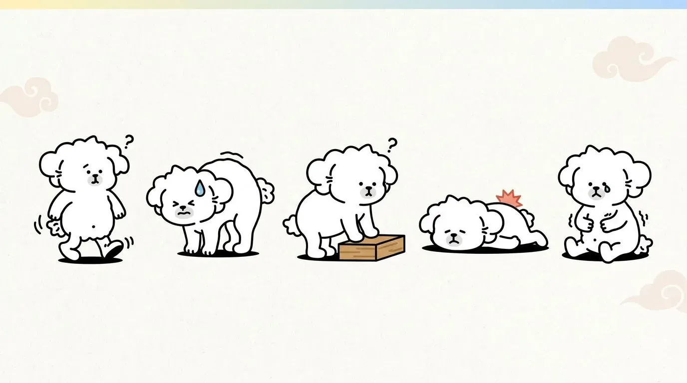
Grade 1-2: Pain and Wobbly Gait (The Critical Window)
Grade 1 is characterized by pain alone — no neurological deficits are present. Your dog is sensitive to touch along the spine, may yelp spontaneously or when moved, and holds an abnormal posture (arched back, lowered head). Motor function is completely intact.
Grade 2 adds proprioceptive ataxia: your dog is ambulatory but walks unsteadily, crossing their legs, stumbling, or appearing “drunk.” Proprioception (the ability to sense where the paws are in space) is mildly impaired, but the dog can still bear weight and walk independently.
Grades 1 and 2 represent the critical treatment window. Most dogs treated promptly with strict rest and medication recover fully. The risk of progression to higher grades — particularly in Type I IVDD — makes this stage one where watchful waiting without veterinary assessment is inadvisable.
Grade 3: Partial Paralysis Begins
Grade 3 means the dog is paretic (partially paralyzed) but still ambulatory. They can walk, but with significant weakness — the hind limbs (in thoracolumbar IVDD) may buckle, knuckle under, or fail to coordinate properly. Deep pain sensation remains intact.
At Grade 3, the cord compression is more substantial. Conservative management can still succeed in some cases, but the success rate drops compared to Grades 1-2, and surgical consultation is typically recommended.
Grade 4-5: Paralysis and Loss of Deep Pain Sensation
Grade 4 is non-ambulatory paraplegia: the dog cannot walk and cannot support their own weight, but still responds to deep pain stimulation in the paws. With prompt surgical decompression, prognosis remains reasonably good for recovery of motor function.
Grade 5 represents complete loss of deep pain sensation in addition to paralysis. Testing deep pain involves applying firm pressure (with a hemostat or similar instrument) to the phalanges — a dog with intact deep pain will react consciously (turning the head, vocalizing). Absence of this response indicates severe spinal cord injury.
Loss of deep pain is a prognostic watershed. Surgery within the first 24-48 hours after deep pain loss gives the best chance of recovery (approximately 50-60% in published case series). Beyond 48 hours, the prognosis for meaningful motor recovery declines significantly, though it is not zero.
| Grade | Neurological Status | Ambulatory | Deep Pain | Typical Treatment Path |
|---|---|---|---|---|
| 1 | Pain only | Yes | Intact | Conservative: rest + medication |
| 2 | Ataxia, mild weakness | Yes (unsteady) | Intact | Conservative; monitor closely |
| 3 | Paresis, significant weakness | Yes (impaired) | Intact | Conservative or surgery (case-dependent) |
| 4 | Paralysis | No | Intact | Surgery strongly recommended |
| 5 | Paralysis + deep pain absent | No | Absent | Emergency surgery; prognosis guarded |
IVDD Symptoms by Spinal Region
The location of disc herniation determines which body parts are affected. Most IVDD occurs in the thoracolumbar region (mid-back), but cervical (neck) and lumbosacral (lower back) disease produce distinct symptom patterns that owners and veterinarians must distinguish.
Cervical (Neck) IVDD: Head Position, Front Leg Signs
Cervical IVDD — typically at C2-C3 or C3-C4 — compresses the cord in the neck region, affecting the front limbs and sometimes all four limbs.
Signs of cervical IVDD:
- Reluctance to turn the head, especially downward toward the food bowl
- Low head carriage; dog holds the nose closer to the ground
- Crying out or yelping when touched on the neck or when moving
- Stiff, stilted front leg gait
- In severe cases: tetraparesis (weakness in all four limbs) or tetraplegia
- Neck muscle spasms (detectable on palpation)
Cervical IVDD is often intensely painful even at low grades, because the cervical spinal canal is relatively small relative to cord diameter. Dogs may scream in pain in the night for no apparent reason — a presentation that frequently alarms owners.
Thoracolumbar (Mid-Back) IVDD: Hind Leg Weakness, Posture Changes
Thoracolumbar IVDD (most commonly T12-T13, T13-L1, or L1-L2) is the most common IVDD location overall, particularly in dachshunds, where it accounts for the majority of cases.
Signs of thoracolumbar IVDD:
- Hunched or arched back (kyphosis) — the dog’s back rises into a “tent” shape
- Reluctance to jump, climb stairs, or use the hind limbs for pushing off
- Hind limb ataxia, weakness, or dragging of the hind paws
- Knuckling: the paw lands on the top surface rather than the pad
- Bladder dysfunction (inability to urinate or urinary leakage) in higher grades
- Fecal incontinence in severe cases
| Symptom | Thoracolumbar IVDD | Cervical IVDD |
|---|---|---|
| Hind limb weakness/paralysis | Yes | No |
| Front limb weakness | Rare (caudal thoracic) | Yes |
| Neck pain signs | No | Yes |
| Bladder/bowel dysfunction | Yes (mid-higher grades) | Rarely |
| Back arching | Yes | Neck stiffness |
Lumbosacral IVDD: Tail, Bladder, and Bowel Signs
Lumbosacral (LS) disc disease affects the junction between the lumbar spine and sacrum (L7-S1). This location is below the spinal cord itself — which ends at approximately L5-L6 in dogs — so compression here injures nerve roots rather than the cord directly (a condition called cauda equina syndrome).
Signs of lumbosacral IVDD:
- Reluctance to wag the tail, or a low-carried, limp tail
- Pain on palpation over the lower back or when lifting the tail
- Hind limb weakness that worsens with exercise, often described as the dog “sitting down” during walks
- Urinary and fecal incontinence (sphincter dysfunction is hallmark)
- Muscle atrophy in the hind quarters over time
- Self-mutilation or excessive licking at the perineal area (referred pain)
LS disease is frequently underdiagnosed because signs are often intermittent and milder at presentation than thoracolumbar IVDD. The absence of dramatic paralysis can lead owners — and sometimes veterinarians — to attribute the signs to arthritis or aging.
Diagnosis: What to Expect at the Vet
If IVDD is suspected, a systematic diagnostic approach establishes both the location and severity of disc herniation, which directly determines treatment decisions.
Neurological Exam and What It Reveals
The neurological examination is the starting point. A veterinarian assesses:
- Gait and posture: ambulatory status, ataxia, knuckling
- Postural reactions: proprioceptive positioning (placing the paw on its dorsal surface — a neurologically intact dog corrects it immediately)
- Spinal reflexes: testing deep tendon reflexes (e.g., patellar reflex) to localize the lesion to a spinal cord segment
- Pain assessment: response to spinal palpation, and deep pain testing at each grade
- Muscle tone and bulk: for signs of lower vs. upper motor neuron disease
Based on the neurological exam alone, a skilled clinician can usually narrow the lesion to a spinal cord region (cervical, thoracic, lumbar, or lumbosacral) before any imaging.
Why MRI Matters More Than X-Ray
Plain radiographs (X-rays) can suggest disc disease — calcified disc material, narrowed disc spaces, or collapsed vertebral endplates are visible signs — but X-rays cannot image the spinal cord itself. A disc can herniate significantly without producing radiographic changes, and calcified material visible on X-ray may not be the disc currently causing compression.
MRI (magnetic resonance imaging) is the gold standard for IVDD diagnosis. It directly visualizes:
- The extent and location of spinal cord compression
- Whether herniation is dorsal, lateral, or circumferential
- The degree of spinal cord signal change (myelomalacia — a grave prognostic sign)
- Concurrent abnormalities (epidural hematoma, other disc protrusions)
CT myelography (CT scan with contrast injected into the spinal fluid) is an alternative when MRI is unavailable and provides excellent anatomical detail, though it requires spinal fluid injection under anesthesia.
The choice of imaging is important: sending a Grade 4 dog home pending a scheduled MRI in three days is not appropriate. Acute, worsening neurological signs warrant same-day or next-day imaging.
Treatment Options: Conservative Care to Surgery
Treatment decisions in IVDD are primarily driven by neurological grade and the trajectory of signs (stable, improving, or deteriorating).
Grades 1-2: Strict Rest, Medication, and Monitoring
For dogs presenting at Grade 1 or 2, conservative management is the first-line approach. It requires two essential components that owners frequently underestimate: the strictness of the rest and the duration.
Strict crate rest means exactly that. The dog lives in a crate or small pen, approximately twice their body length, for 4-6 weeks. No jumping, no stairs, no running — even brief periods of unsupervised activity can re-herniate or worsen an unstable disc. Leash walks are limited to toileting only, kept brief and at a slow pace.
Medications typically prescribed:
- NSAIDs (non-steroidal anti-inflammatory drugs): reduce inflammation around the disc and cord
- Corticosteroids: sometimes used for acute severe cases, though concurrent NSAID use is contraindicated
- Muscle relaxants (e.g., methocarbamol): address paraspinal muscle spasm
- Gabapentin: neuropathic pain management, increasingly used as an adjunct
Close monitoring during conservative management is essential. If signs worsen — particularly if a Grade 1-2 dog deteriorates to Grade 3 or beyond — the treatment plan must be reassessed urgently. Any progression to loss of motor function or bladder control is a sign that conservative management is failing.
Grades 3+: When Surgery Becomes Necessary
Surgical decompression — most commonly a hemilaminectomy for thoracolumbar IVDD or ventral slot for cervical IVDD — removes the herniated disc material from the spinal canal, eliminating the source of compression.
The decision to pursue surgery considers:
- Neurological grade (Grade 3 with failed conservative management, Grade 4-5 regardless)
- Rate of deterioration (rapid progression is a surgical emergency)
- Duration of signs (delayed surgery for Grade 4-5 cases worsens prognosis)
- Owner factors (surgical access, cost, post-operative care capacity)
Success rates by grade (surgery):
- Grade 2-3: 95%+ recovery of ambulatory function
- Grade 4: approximately 85-90% with prompt surgery
- Grade 5 with deep pain present <48 hours: approximately 50-60%
- Grade 5 with deep pain absent >48 hours: approximately 25-50%, highly variable
These figures represent return to ambulatory function, not necessarily complete normalization.
Complementary Therapies: Physical Rehab, Laser, and NIR Therapy
Regardless of whether treatment is conservative or surgical, physical rehabilitation is an evidence-based component of recovery that significantly improves outcomes, particularly for Grades 3-5.
Hydrotherapy (underwater treadmill): Allows the dog to exercise limbs in a reduced-gravity environment. Water’s buoyancy supports body weight while resistance builds muscle strength. Studies document faster recovery of ambulatory function with hydrotherapy in post-surgical IVDD dogs.
Laser therapy (low-level laser / photobiomodulation): Class IV therapeutic lasers deliver wavelengths in the 810-980 nm range that penetrate soft tissue, promoting mitochondrial activity, reducing neuroinflammation, and improving local circulation. Multiple published studies in small animal neurology support laser therapy as an adjunct for spinal cord injury recovery.
Near-infrared (NIR) therapy: Near-infrared wavelengths (700-1200 nm) penetrate deeper into tissue than visible light, reaching paraspinal muscles, nerve tissue, and periosteal structures. NIR therapy targets the same photobiomodulation pathways as laser therapy, promoting ATP synthesis in recovering nerve cells and reducing inflammatory cytokines. For dogs with chronic IVDD pain or those in rehabilitation, NIR therapy applied to the paraspinal musculature addresses both the pain component and the tissue-healing environment. The near-infrared therapy for dogs article covers the mechanisms and application protocols in detail.
Acupuncture: Veterinary acupuncture has emerging evidence for spinal cord injury, with proposed mechanisms including endorphin release, local anti-inflammatory effects, and modulation of spinal cord pain pathways. Some veterinary neurologists integrate acupuncture into the post-surgical recovery protocol, particularly for Grade 4-5 patients where every additional recovery tool is valuable.
Electrical stimulation (NMES/TENS): Neuromuscular electrical stimulation can be used to maintain muscle bulk and promote re-innervation in paretic limbs during the recovery phase.
Home Care for Dogs with IVDD
Whether your dog is in conservative management or post-surgical recovery, the home environment critically affects outcomes.
Setting Up a Safe Recovery Environment
The goal is to eliminate opportunities for sudden movement, jumping, or impact while maintaining comfort and hygiene.
Crate setup checklist:
- Wire or plastic crate approximately 2x the dog’s length — large enough to stand and turn, not so large that the dog can build momentum
- Thick, supportive bedding (orthopedic foam or memory foam) to protect pressure points
- Water accessible without requiring the dog to reach or stretch
- Non-slip mat under the crate and anywhere the dog will walk — smooth floors are a significant fall hazard for ataxic dogs
- Baby gates blocking stair access even if the crate door is open
- Ramps (not steps) to get on and off furniture if your vet has permitted limited access
For paralyzed dogs requiring bladder expression, set up the expression area with washable, absorbent pads. Keep cleaning supplies immediately accessible — a paralyzed dog in a soiled environment risks pressure sores and urinary tract infection.
How to Lift and Carry Your Dog Safely
Improper lifting is one of the most common ways owners inadvertently worsen IVDD. The spine must be supported in neutral alignment throughout the lift.
For thoracolumbar IVDD: Place one hand under the chest (cranial to the front legs), the other hand under the abdomen or hindquarters. Lift as a single horizontal unit — do not allow the back to sag. Avoid lifting from the front legs alone or by the scruff.
For cervical IVDD: Support the head and neck in alignment with the body. Do not flex or extend the neck during the lift. Both hands should support the thorax while the head rests naturally on the forearm.
For dogs weighing more than 10-15 kg, a supporting sling under the abdomen (a rolled towel or commercial cart harness) assists with mobility without requiring the dog to bear full weight.
Exercise and Activity: What’s Allowed at Each Stage
Activity restrictions must be communicated clearly by the attending veterinarian based on grade and progress. The general framework:
| Recovery Phase | Allowed Activity | Duration |
|---|---|---|
| Acute rest (Weeks 1-4) | Leash toileting only, 2-3 min max | 4-6 weeks |
| Gradual reintroduction (Weeks 5-8) | Short leash walks on flat, non-slip surfaces | Building to 5-10 min 3x/day |
| Rehabilitation phase | Structured rehab exercises (as guided by veterinary physiotherapist) | Ongoing |
| Maintenance phase | Regular, controlled exercise; no high-impact activity | Permanent lifestyle modification |
High-impact activities — jumping off furniture, running freely, rough play — should be permanently reduced or eliminated in chondrodystrophic breeds after any IVDD episode, given the recurrence risk. Harnesses (rather than collars) are standard for all walks to avoid any spinal stress from leash jerking.
Keeping your dog at a healthy body weight is one of the most effective long-term management strategies. Excess weight increases compressive loads on the already vulnerable discs and accelerates degeneration — an issue discussed further in the context of senior dog joint care.
When differentiating IVDD from other causes of hind limb weakness — such as patellar luxation — the pattern of onset is often the key clue: IVDD typically produces more acute neurological signs, while patellar luxation presents as an intermittent mechanical lameness without spinal pain. Similarly, the gradual joint stiffness associated with dog arthritis symptoms differs from the acute pain and neurological deterioration characteristic of disc herniation, though both conditions can coexist in older dogs.
FAQ
What are the first signs of IVDD in dogs?
Can a dog recover from IVDD without surgery?
What is the final stage of IVDD in dogs?
How long does IVDD recovery take?
What is the life expectancy of a dog with IVDD?
Related Articles
Dog Arthritis: Symptoms and Management
Recognize early signs of arthritis in dogs and learn effective management strategies for pain relief and improved mobility.
The Hidden Link Between Your Dog's Weight and Joint Health
Extra weight does far more than strain your dog's joints — it actively inflames them. Learn the dual-pathway science and a practical roadmap to protect joint health.
Complete Guide to Patellar Luxation in Dogs
Learn about the causes, symptoms, grades, treatment options, and prevention of patellar luxation in dogs.