If Your Dog's Hind Legs Are Getting Thinner, It Could Be Sarcopenia
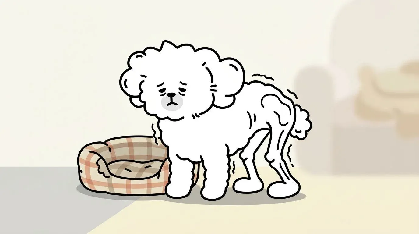
You notice something one evening while your dog is walking away from you. The hind legs look thinner than they used to. The haunches seem narrower, the thighs less full. Your dog moves fine, more or less — so you chalk it up to old age.
That observation deserves more attention than it usually gets. What you are seeing is likely muscle atrophy, and in senior dogs, it is often the earliest visible sign of sarcopenia — the progressive, age-associated loss of muscle mass that affects the majority of dogs over seven years old. Unlike the dramatic collapse we associate with serious illness, sarcopenia advances quietly, one missed walk at a time, until the consequences become impossible to ignore.
This guide explains what dog muscle atrophy is, why it happens, how to assess it at home using a validated scoring system, and what the current evidence supports for recovery.
What Is Muscle Atrophy in Dogs?
Muscle atrophy is the reduction in muscle mass and fiber size. In everyday terms: the muscles shrink. It is not a disease on its own but a physiological response to one or more underlying processes — insufficient use, inadequate nutrition, nerve damage, or systemic illness.
Why Muscles Matter More Than You Think
It is tempting to think of muscles purely as the machinery of movement. They are far more than that. In dogs, skeletal muscle tissue accounts for roughly 40–50% of total body mass and functions as a metabolic organ in its own right.
Muscles serve as the body’s primary reservoir of amino acids, which are mobilized during illness, injury, or caloric deficit to support immune function and tissue repair. They play a central role in glucose metabolism — contracting muscle is one of the largest sites of glucose uptake in the body, and the loss of muscle mass is directly associated with impaired glucose regulation. Muscles also generate heat, contributing to thermoregulation, which matters more for thin or senior dogs in cold climates.
When muscle mass falls, these systemic functions are compromised alongside the more visible loss of strength and mobility.
Disuse Atrophy vs. Neural Atrophy
Not all muscle atrophy in dogs works the same way, and the distinction matters clinically.
Disuse atrophy develops when a muscle is not used adequately. This can happen because of reduced activity (a dog that is largely sedentary), pain-induced limb sparing (a dog that avoids bearing weight on an arthritic or injured leg), or prolonged immobilization after surgery. Disuse atrophy is typically gradual, affects the muscles of a specific limb or region, and — importantly — is largely reversible with appropriate rehabilitation.
Neural atrophy occurs when the nerve supply to a muscle is damaged or interrupted. Spinal cord compression (from intervertebral disc disease, for example), nerve root damage, or peripheral neuropathy can all cause the denervated muscles to waste rapidly — within days to weeks rather than months. Neural atrophy progresses faster and recovers more incompletely than disuse atrophy, and it requires urgent veterinary evaluation.
The practical difference for owners: disuse atrophy tends to be symmetrical or gradual and correlates with behavioral changes like reduced activity. Neural atrophy often appears suddenly, may affect one limb disproportionately, and is frequently accompanied by obvious neurological signs — weakness, knuckling, loss of deep pain sensation.
What Causes Muscle Loss in Dogs?
Dog muscle atrophy does not have a single cause. In practice, multiple factors often compound each other.
Aging and Sarcopenia — The 15–20% Rule After Age 7
Sarcopenia (from the Greek sarx, flesh, and penia, loss) is the age-associated decline in skeletal muscle mass, strength, and function. It is not an aberration — it is the default trajectory of aging muscle in all mammals, including dogs.
Research in geriatric dogs has documented muscle mass losses in the range of 15–20% between ages 7 and 12, even in otherwise healthy animals with adequate nutrition and activity. The mechanisms involve a shift in the balance between muscle protein synthesis and breakdown: synthesis slows with age, and the anabolic response to protein intake becomes blunted — meaning older muscles respond less efficiently to the same nutritional signals that would readily stimulate growth in a younger animal.
Large breed dogs — Labrador Retrievers, German Shepherds, Golden Retrievers, Bernese Mountain Dogs — often show clinically meaningful sarcopenia earlier than small breeds, partly because of their greater total muscle mass and faster overall aging trajectory.
Inactivity and Sedentary Lifestyle
Muscle is metabolically expensive tissue. The body maintains it only when it is regularly used. When a dog becomes sedentary — due to a low-activity lifestyle, weather, pain, or an owner’s schedule — muscle protein breakdown begins to outpace synthesis within days. The hind limb muscles, which are among the largest in the body, are often the first to show visible reduction.
This is where recognizing early signs of decreased activity matters: a dog that is walking less, sleeping more, and declining stairs may be losing muscle mass even before the change is visible to the naked eye. Inactivity is both a cause and a consequence of muscle loss — a cycle that is difficult to break once established.
Nutritional Deficiency — Protein and Key Amino Acids
Muscle protein synthesis requires adequate dietary protein and, specifically, adequate essential amino acids. Leucine — a branched-chain amino acid (BCAA) — plays an outsized role as an anabolic signal: it directly activates the mTORC1 pathway, the molecular switch that turns on muscle protein synthesis. Research in dogs and other mammals has demonstrated that leucine supplementation alone can stimulate muscle protein synthesis even without changes to total protein intake.
Dogs fed low-protein diets (below the WSAVA’s recommended minimums), or diets high in poor-quality protein sources that are low in essential amino acids, are at elevated risk for accelerated muscle loss. This is particularly relevant for senior dogs, whose protein requirements are, if anything, higher than those of young adults — not lower, as was once assumed. For a broader view of how diet affects musculoskeletal health, the guide to joint-supportive foods for dogs covers the overlap between anti-inflammatory nutrition and muscle preservation.
Chronic Disease and Cachexia
Several systemic diseases accelerate muscle wasting through mechanisms beyond simple disuse or protein deficiency. This form of pathological muscle loss is called cachexia, and it is distinct from sarcopenia in important ways.
Cachexia is driven primarily by inflammation — elevated circulating cytokines (TNF-α, IL-6, IL-1β) that directly promote muscle protein breakdown while simultaneously suppressing appetite. Chronic diseases commonly associated with cachexia in dogs include congestive heart failure, chronic kidney disease (CKD), cancer, and inflammatory bowel disease.
The prognostic distinction matters: cachexia, when caused by an active inflammatory disease process, tends to progress faster and responds less completely to nutritional and exercise interventions than sarcopenia or disuse atrophy. Treatment focuses on addressing the underlying disease. That said, recent veterinary nutrition research has revised older thinking about protein restriction in CKD: for dogs in early to moderate CKD stages (IRIS Stages 1–3), maintaining adequate protein intake is now understood to be critical for muscle preservation, as excessive restriction can accelerate muscle wasting without meaningfully slowing disease progression.
Dogs with chronic pain from arthritis or joint conditions frequently develop secondary muscle atrophy through a combination of disuse (sparing painful limbs) and low-grade systemic inflammation — illustrating how the same dog can experience multiple overlapping mechanisms simultaneously.
How to Spot Muscle Atrophy: Signs by Stage
Dog muscle atrophy in hind legs is the most commonly noticed finding by owners, but atrophy can affect any major muscle group — the epaxial muscles along the spine, the hindquarters, the shoulder girdle, or the temporalis muscles of the jaw.
Early Stage — Reduced Thigh and Hip Muscle Tone
At the earliest stage, muscle loss is subtle enough that owners often mistake it for normal variation or seasonal weight fluctuation. The thighs and gluteal muscles may appear slightly less rounded. Running your hands along the dog’s spine, you may notice the vertebral processes feel more prominent than before, though a layer of tissue still covers them.
The dog’s behavior may offer earlier clues than appearance: slight reluctance to jump or climb stairs, a preference for lying on one side, or brief hesitation before rising from rest. Dogs experiencing pain-related behavioral changes often self-limit activity in ways that accelerate the very muscle loss that will worsen their pain.
Moderate Stage — Difficulty Climbing Stairs, Hind Leg Wobble
As atrophy progresses, functional consequences become apparent. The dog may struggle on stairs — pausing mid-way, taking them slowly, or skipping them entirely. Jumping into the car becomes unreliable. On flat ground, the hind legs may occasionally wobble or bunny-hop rather than alternate normally.
Visually, the hind limbs now look noticeably thinner compared to the forequarters. The pelvis appears more prominent. In some dogs, particularly large breeds, the topline (the line of the back seen from the side) begins to appear slightly dipped or uneven.
Advanced Stage — Sunken Topline, Inability to Rise
In severe muscle atrophy, the dog struggles to rise from a lying position without assistance. The topline is markedly sunken as the epaxial muscles along the spine waste. Hind leg weakness means the dog may cross its legs, stumble, or collapse during standing. The temporal muscles of the face may appear hollowed, giving the head a gaunt appearance.
At this stage, muscle mass loss is visible on the ribcage and shoulders as well. If the dog has also lost body fat — as seen in cachexia — the ribs and hip bones are prominently visible. This is a medical emergency that warrants immediate veterinary evaluation.
Self-Assessment with BCS and MCS Scoring
Two scoring systems help owners and veterinarians track muscle and body condition objectively.
Body Condition Score (BCS) assesses overall fat reserves on a 1–9 scale (or 1–5 in some systems). It focuses on rib palpability, waist definition, and abdominal tuck. A BCS of 4–5/9 is ideal. The limitation of BCS for this discussion is that it does not directly measure muscle — a dog can have a normal BCS while losing significant muscle mass beneath adequate fat cover.
Muscle Condition Score (MCS) is designed specifically to assess skeletal muscle and is recommended by the World Small Animal Veterinary Association (WSAVA). It uses a four-point scale:
| MCS Grade | Description | What You Feel |
|---|---|---|
| Normal | Adequate muscle mass over spine, scapulae, skull, and pelvis | Bones covered with a firm layer of muscle |
| Mild loss | Mild muscle atrophy over at least one area | Vertebrae and pelvis slightly more prominent |
| Moderate loss | Atrophy over multiple areas | Bones clearly palpable; muscle tissue feels thin |
| Severe loss | Marked atrophy throughout | Bones prominently visible; minimal covering tissue |
How to do a basic MCS at home: Run both hands firmly along your dog’s spine, pressing gently but with purpose. Then feel over the tops of the shoulder blades, the hip bones, and — in medium to large dogs — the base of the skull behind the ears. Note which bony landmarks feel prominent or sharp. Then compare the circumference of both thighs by encircling each with your hands at the same point — a measurable difference of 1 cm or more between sides suggests asymmetric muscle loss worth investigating.
Treatment and Management Strategies
The question dog owners most want answered is whether muscle atrophy in dogs can be reversed. For disuse atrophy and sarcopenia, the answer is yes — partially and meaningfully, with the right approach.
Nutrition — High-Quality Protein and Key Supplements (Leucine, Omega-3, Vitamin D)
Nutrition is the foundation of muscle preservation and recovery. Three principles matter most.
Adequate total protein: The WSAVA recommends a minimum of 18% protein on a dry matter basis for adult dogs, but senior dogs with muscle loss benefit from higher intakes — in the range of 25–30% dry matter — provided they do not have contraindications such as advanced kidney disease. Protein quality matters as much as quantity: look for whole-meat protein sources (chicken, beef, salmon, eggs) that are rich in essential amino acids rather than plant-based proteins that may be deficient in specific amino acids.
Leucine: Among all amino acids, leucine has the strongest evidence as a direct stimulant of muscle protein synthesis, independent of total protein intake. Some clinical veterinary nutritionists recommend leucine-enriched foods or leucine supplementation for dogs with confirmed sarcopenia. Leucine is found naturally in high concentrations in eggs, chicken breast, and beef.
Omega-3 fatty acids (EPA and DHA): Fish oil-derived omega-3s reduce inflammatory cytokine activity — the same cytokines that drive muscle breakdown in cachexia and in chronic inflammatory conditions. Beyond their anti-inflammatory role in joints (covered in the omega-3 and joint health guide), EPA and DHA have been shown to preserve lean body mass in animal models of sarcopenia.
Vitamin D: Vitamin D deficiency is associated with reduced muscle fiber size and impaired neuromuscular function across species. Some senior dogs are deficient, particularly those with limited sun exposure. Testing and, if necessary, supplementation is worth discussing with a veterinarian, especially in dogs with confirmed muscle loss.
For a comprehensive look at how food choices support musculoskeletal health alongside supplementation, the joint-health food guide provides practical dietary frameworks that apply equally to muscle preservation.
Exercise Programs — Short, Frequent, Low-Impact
Exercise is the most powerful stimulus for muscle protein synthesis — but for dogs with existing atrophy, the approach must be graduated carefully.
The principle is simple: muscles grow in response to load. Without the mechanical stimulus of weight-bearing and resistance, nutritional strategies have limited effect. With appropriate exercise, even significantly atrophied muscles can respond.
For early to moderate atrophy:
- Two to three short leash walks daily (10–15 minutes each) are more effective than a single long walk
- Walking on slightly uneven surfaces (grass, gentle hills) engages more stabilizer muscles than flat pavement
- Slow, controlled sitting and standing exercises (“dog squats” — asking the dog to sit and stand from a sit repeatedly) build hind limb strength specifically
- Cavaletti poles — low bars spaced at the dog’s stride length — encourage deliberate foot placement and hind leg engagement
For advanced atrophy or dogs with pain:
- Start with supported standing: allow the dog to stand while you support the hindquarters with a towel or sling
- Even brief periods of weight-bearing (2–3 minutes several times daily) provide meaningful stimulus
- Avoid high-impact activities, sudden acceleration or deceleration, and rough play until baseline strength is re-established
One common mistake: resting a dog completely because it seems tired or in pain. Rest allows existing atrophy to worsen. The goal is movement that is appropriate in duration and intensity — not inactivity. Maintaining a consistent, gentle routine matters more than individual session intensity.
Physical Therapy Options — Hydrotherapy, NIR Therapy, Massage
For dogs with significant atrophy, especially when pain limits normal exercise, formal rehabilitation therapies substantially accelerate recovery.
Hydrotherapy (underwater treadmill or pool swimming) is the gold standard rehabilitation modality for canine muscle atrophy. Water supports a significant portion of the dog’s body weight, allowing full range-of-motion movement with greatly reduced joint loading. The resistance of water simultaneously provides a meaningful muscular challenge. Dogs that cannot bear full weight on land can often walk on an underwater treadmill for extended periods, providing the repetitive muscular stimulus that drives recovery. Multiple studies in veterinary rehabilitation literature support hydrotherapy for rebuilding hind limb muscle mass after orthopedic surgery, spinal cord injury, and sarcopenia.
Near-infrared (NIR) therapy works through a different mechanism. NIR light (wavelengths 700–1100 nm) penetrates soft tissue and is absorbed by cytochrome c oxidase in mitochondria, stimulating ATP production and increasing local microcirculation. In the context of muscle atrophy, improved blood flow means better oxygen and nutrient delivery to recovering muscle fibers. NIR therapy also modulates inflammatory mediators — reducing the cytokine activity that drives muscle breakdown in chronic conditions. For dogs managing concurrent joint pain alongside muscle loss, home NIR therapy offers a practical adjunct that owners can apply consistently between clinic visits.
Therapeutic massage improves local circulation, reduces muscle tension, and may help counteract the disuse-associated stiffness that makes dogs reluctant to move. It also provides an opportunity for owners to monitor muscle tone changes over time. Massage is not a substitute for active exercise but works well as a complement.
Breaking the Vicious Cycle of Muscle Loss
One of the more important concepts in managing dog muscle atrophy is understanding that it is self-reinforcing once established.
The cycle works like this: muscle weakness makes movement effortful or painful → the dog moves less → reduced movement accelerates muscle loss → weakness increases → and so on. Simultaneously, weakened muscles no longer adequately stabilize and support joints → joint stress increases → joint pain develops or worsens → the dog avoids using painful limbs → more atrophy follows.
This is why dogs with arthritis-related muscle wasting and joint pain often deteriorate faster than the arthritis alone would predict. The muscle and joint problems feed each other. And it is why intervention earlier in the cycle — when atrophy is mild and pain is manageable — produces far better outcomes than waiting until the dog can no longer rise unassisted.
Managing body weight is also part of cycle-breaking. Obesity imposes additional mechanical load on already-weakened muscles and joints, accelerating both the pain and the disuse. Conversely, underweight dogs lack the nutritional reserves for muscle repair. The relationship between weight and joint health explains these connections in detail.
For a whole-body approach to managing the aging dog — including environmental modifications, exercise pacing, and the integration of supportive therapies — the comprehensive senior dog joint care guide provides a practical framework that addresses muscle and joint health together.
When to See a Veterinarian
Not all muscle atrophy warrants the same urgency. But certain findings call for prompt professional evaluation.
Seek veterinary care promptly if:
- Muscle loss appears rapidly (over days to weeks rather than months) — this pattern suggests neural atrophy or cachexia rather than sarcopenia
- One limb is losing muscle significantly faster than the other, especially if accompanied by knuckling, dragging, or loss of coordination
- Your dog cannot rise from the floor without assistance, or collapses under its own weight
- Appetite has decreased significantly alongside the muscle loss — this suggests systemic illness driving cachexia
- You notice hollowing of the temporal muscles of the face (behind the eye socket) — masticatory muscle myositis is a specific immune-mediated condition requiring targeted treatment
- Your dog is losing weight rapidly even with maintained or increased food intake
Expect the veterinarian to assess:
- Neurological exam (reflexes, conscious proprioception, pain response) to rule out neural atrophy
- Blood and urine panels to screen for underlying disease (kidney function, thyroid, liver, diabetes, inflammatory markers)
- Muscle Condition Score and Body Condition Score
- In some cases, radiographs to assess spinal or joint changes contributing to disuse
- Discussion of dietary protein adequacy based on the dog’s current food and health status
Early diagnosis is what separates a reversible condition from a manageable but progressive one. The earlier interventions begin — nutritional adjustment, graduated exercise, supportive therapies — the more muscle function can be preserved or restored.
FAQ
Can a senior dog regain muscle mass?
Is muscle atrophy in dogs painful?
What supplements help with dog muscle atrophy?
What is the life expectancy of a dog with muscle atrophy?
How do I check my dog's muscle condition at home?
Related Articles
The Hidden Link Between Your Dog's Weight and Joint Health
Extra weight does far more than strain your dog's joints — it actively inflames them. Learn the dual-pathway science and a practical roadmap to protect joint health.
Dog Arthritis: Symptoms and Management
Recognize early signs of arthritis in dogs and learn effective management strategies for pain relief and improved mobility.
Complete Guide to Patellar Luxation in Dogs
Learn about the causes, symptoms, grades, treatment options, and prevention of patellar luxation in dogs.
Is Your Dog Showing Signs of IVDD? Symptoms, Stages, and What to Do
Learn to recognize IVDD in dogs symptoms by grade and spinal region, understand treatment options from conservative care to surgery, and manage recovery at home.
Dog Cruciate Ligament Tear: 7 Things Every Owner Must Know
Dog cruciate ligament tear guide: recognize symptoms, compare TPLO vs TTA surgery, follow a phased recovery timeline, and protect the opposite knee.
Dog Hip Dysplasia: 6 Essential Facts Every Owner Should Know
Learn the key facts about dog hip dysplasia — from at-risk breeds and early symptoms to PennHIP vs OFA diagnosis and home care exercises.
Why Is My Dog Limping? A Cause-by-Cause Diagnosis Guide
Dog limping on front or back leg? Learn the most common causes by leg and age, a home observation checklist, and when the limp needs emergency care.