 Cirius Petpedia
 Veterinary Guide to Vestibular Disease in Dogs: Symptoms, Home Care, and Recovery
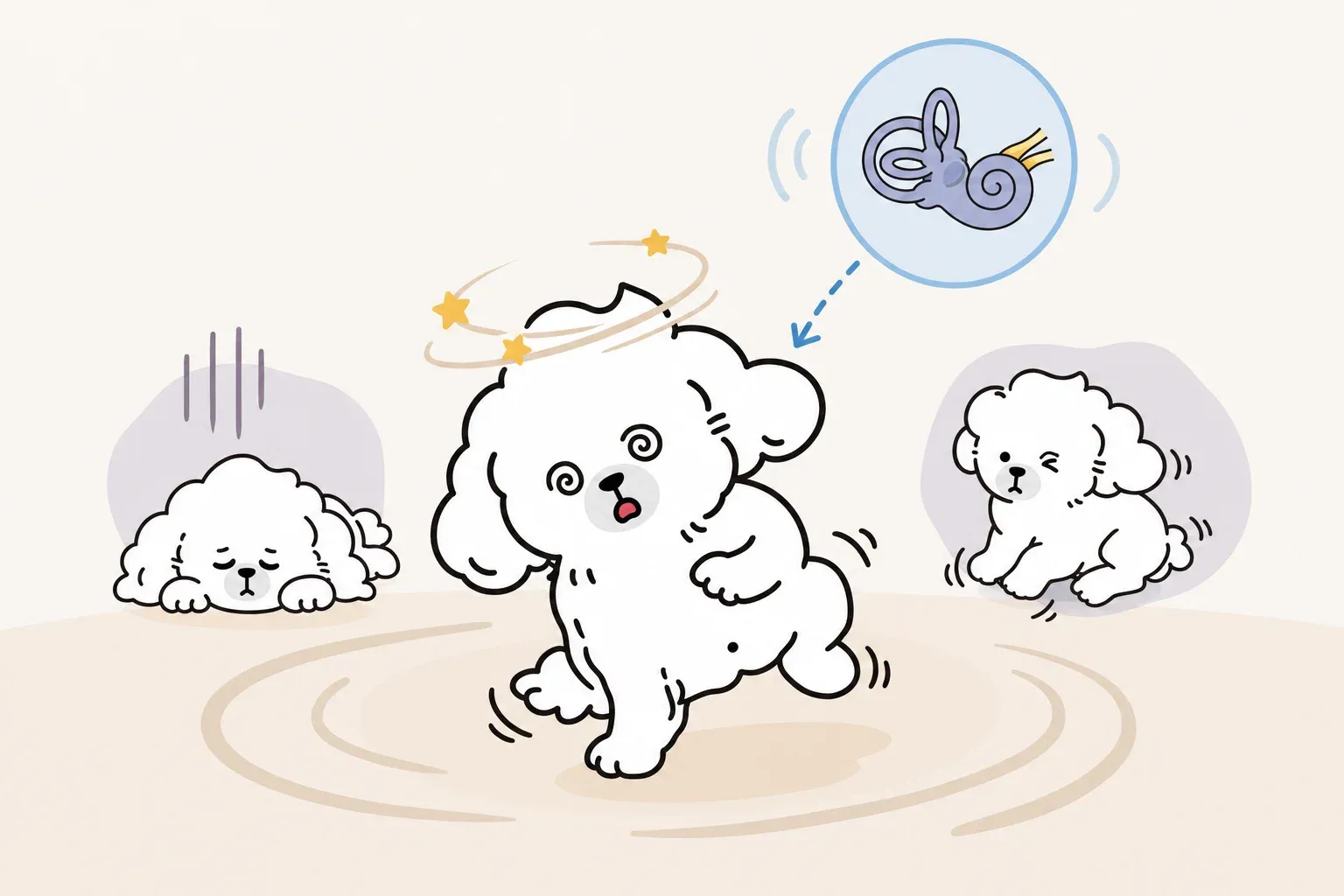
Your dog woke up unable to walk straight. His head is tilted to one side, his eyes are darting back and forth, and he’s fallen over trying to stand. Your first instinct is the worst — stroke, brain tumor, something catastrophic.
In many cases, it isn’t. What you’re likely witnessing is dog vestibular disease, one of the most dramatic-looking yet often self-resolving conditions in veterinary medicine. Understanding what it is, how to tell the dangerous form from the benign one, and how to support your dog through recovery can make a frightening situation far more manageable.
What Is Vestibular Disease in Dogs?
The vestibular system is the body’s balance control center. It processes signals about head position and movement, then coordinates the eyes, limbs, and trunk to keep the body upright and moving smoothly. When this system malfunctions in dogs, the result is sudden, pronounced disorientation — often so severe that owners mistake it for a stroke.
How the Vestibular System Works
The vestibular apparatus consists of two anatomical components working in tandem. The peripheral component — the inner ear and the vestibular branch of the eighth cranial nerve — detects motion and gravitational changes. The central component — primarily the brainstem and cerebellum — integrates those signals and issues corrective commands to the muscles.
When either component is disrupted, the brain receives faulty or absent balance information. The resulting clinical signs reflect the brain’s attempt to compensate for input that no longer makes sense: the eyes dart involuntarily (nystagmus), the head tilts toward the affected side, and the limbs struggle to coordinate. For an owner watching this unfold in real time, it looks terrifying.
Peripheral vs Central Vestibular Disease
The most important distinction in vestibular disease is anatomical: where is the problem located?
| Feature | Peripheral | Central |
|---|---|---|
| Location | Inner ear / cranial nerve VIII | Brainstem / cerebellum |
| Common causes | Idiopathic syndrome, ear infection, ototoxic drugs | Tumor, inflammatory disease, stroke, trauma |
| Nystagmus direction | Horizontal or rotary | Vertical or changing direction |
| Mental alertness | Normal | Often depressed or altered |
| Postural reactions | Usually normal | Frequently abnormal |
| Prognosis | Generally excellent | Depends on underlying cause |
| Urgency | Can observe for 24–48 hours | Requires immediate evaluation |
Peripheral vestibular disease accounts for the majority of cases and carries a favorable prognosis. Central vestibular disease is less common but far more medically serious. Distinguishing between the two is the first job of your veterinarian — and the reason a vet visit is always warranted on first presentation.
Recognizing the Symptoms of Dog Vestibular Disease
The clinical picture of vestibular disease tends to appear suddenly, often seemingly overnight. Symptoms can be alarming in their intensity, particularly at onset.
Head Tilt and Nystagmus
A persistent head tilt — where the dog holds its head cocked to one side — is the hallmark sign. The tilt typically points toward the affected side and may remain even after other symptoms resolve.
Nystagmus (involuntary rapid eye movement) is equally characteristic. The eyes flick rhythmically in one direction — most often horizontal or rotary in peripheral disease. If the nystagmus is vertical (eyes moving up and down) or changes direction, that is a red flag for central involvement. The technical term for the slow phase of the nystagmus pointing away from the lesion side is called the “fast phase” pointing toward the lesion — a detail your vet will assess to localize the lesion.
Loss of Balance, Circling, and Vomiting
Dogs with vestibular disease often lose the ability to walk in a straight line. They may lean heavily to one side, circle in a tight arc, or fall over repeatedly when attempting to stand. This is called ataxia (incoordination), and it can range from a subtle wobble to complete inability to rise.
Nausea and vomiting are common secondary effects, caused by the same mechanism as motion sickness in humans — the brain is receiving conflicting signals about motion and position. Many dogs refuse food during the acute phase.
Symptom Severity: A Self-Assessment Checklist
Use this checklist to assess your dog’s condition before calling your vet:
Immediate emergency — call an emergency clinic now:
- Cannot stand or rise at all
- Eyes flicking vertically (up and down) or changing direction
- Significantly depressed, unresponsive, or stuporous
- Seizure activity occurring alongside balance loss
- Symptoms worsening rapidly over hours rather than stabilizing
- Known head trauma preceding symptoms
Urgent — schedule same-day or next-morning appointment:
- Head tilt present with moderate loss of balance
- Horizontal or rotary nystagmus present
- Vomiting repeatedly but dog is alert and responsive
- Refusing food and water for more than 12 hours
- Symptoms unchanged or slightly worsened after 24 hours
Monitor and call your vet:
- Mild head tilt, dog can still stand and walk
- Occasional stumbling but alert and responsive
- Eating and drinking, even if reduced
- Symptoms visibly improving over the first 24–48 hours
This checklist is a triage tool, not a diagnostic substitute. Any new episode of sudden balance loss deserves veterinary evaluation.
Common Causes of Vestibular Disease in Dogs
Idiopathic Vestibular Syndrome (Most Common in Senior Dogs)
The most frequent diagnosis in middle-aged to senior dogs is idiopathic vestibular syndrome — vestibular dysfunction with no identifiable underlying cause. It is also called “old dog vestibular disease” because of its strong association with dogs over 8 years of age, though it can occur at any age.
The exact mechanism remains unknown, which is what “idiopathic” means by definition. What is known is that the syndrome is self-limiting: the vast majority of affected dogs recover fully or near-fully within 1 to 3 weeks without specific treatment.
Breed data from BMC Veterinary Research and clinical case series suggest that large-breed dogs — German Shepherds, Labrador Retrievers, and Golden Retrievers in particular — appear in vestibular disease case reports at disproportionate rates compared to the general hospital population, though controlled prevalence studies remain limited. Clinicians anecdotally note higher presentation rates in larger, older dogs, which may reflect both genetic predisposition and the longer lifespans now seen in well-cared-for large breeds.
Middle and Inner Ear Infections
Ear infections (otitis media/interna) are the second most common cause of peripheral vestibular disease. When bacterial or fungal infection spreads from the external ear canal into the middle or inner ear, it can inflame or damage the vestibular apparatus directly.
Signs that suggest infection as the underlying cause include a history of chronic ear infections, visible ear discharge, head shaking before vestibular signs appeared, and pain on palpation of the ear. Diagnosis typically requires otoscopic examination, culture of ear secretions, and sometimes CT imaging to assess the tympanic bulla.
Treatment — typically a prolonged course of antimicrobials, often 6 to 8 weeks — can dramatically improve or resolve vestibular signs in these cases. This distinction matters because untreated infectious vestibular disease will not self-resolve the way idiopathic disease does.
Hypothyroidism, Ototoxic Medications, and Tumors
Several less common causes complete the differential:
Hypothyroidism is associated with vestibular dysfunction in dogs, likely through peripheral neuropathy affecting cranial nerve VIII. The link between hypothyroidism and vestibular signs is well-documented, and thyroid supplementation often produces marked improvement. A routine thyroid panel (T4, freeT4, TSH) should be part of the initial workup in middle-aged to older dogs presenting with vestibular disease.
Ototoxic medications — aminoglycoside antibiotics (gentamicin, amikacin), certain diuretics (furosemide at high doses), and some antiseptic ear flushes — can cause direct damage to the sensory hair cells of the inner ear. If your dog has recently received topical or systemic medications of these classes, inform your vet immediately.
Tumors (nasopharyngeal polyps in cats, though rare in dogs; nerve sheath tumors; skull base tumors) can compress the vestibular nerve or disrupt the brainstem, causing progressive vestibular signs. Unlike the sudden onset of idiopathic disease, tumor-related signs often develop gradually and may be accompanied by other neurological deficits.
Is It Vestibular Disease or Something Else?
Vestibular Disease vs Disc Disease vs Stroke: Comparison Table
One of the most important clinical challenges — and the most common source of owner anxiety — is distinguishing vestibular disease from other conditions that cause sudden neurological signs.
| Feature | Vestibular Disease | Disc Disease (IVDD) | Stroke (FCE/Hemorrhagic) |
|---|---|---|---|
| Onset | Sudden (minutes to hours) | Hours to days | Sudden (seconds) |
| Primary sign | Head tilt, nystagmus, circling | Back or neck pain, limb weakness | One-sided weakness or paralysis |
| Pain present? | Typically absent | Often yes (spinal pain) | Typically absent |
| Consciousness | Normal (peripheral) | Normal | Normal to depressed |
| Progression | Stabilizes within 24–72h | Variable (can worsen) | Stabilizes within minutes |
| Nystagmus | Common | Absent | May be present |
| Limb deficits | Gait ataxia only | Weakness, paralysis | Hemiparesis (one side) |
| Key diagnostic test | Neuro exam, MRI if central suspected | Spinal MRI or CT | MRI (diffusion-weighted) |
| Prognosis | Usually excellent | Depends on grade | Variable |
Vestibular disease and disc disease can both cause sudden-onset ataxia, but spinal pain is a key distinguishing feature: most dogs with disc herniation show signs of discomfort on neck or back palpation, while vestibular dogs typically do not. Similarly, a dog losing balance suddenly during what appears to be a seizure episode will have altered consciousness and involuntary motor movements that differ from vestibular ataxia.
Red Flags That Require Emergency Care
Certain features should prompt immediate emergency evaluation, regardless of how the overall picture looks:
- Vertical nystagmus: Eyes moving up-down rather than side-to-side strongly suggests central (brainstem) disease
- Altered mentation: Confusion, stupor, or unresponsiveness points to central involvement
- Rapid progression: Signs worsening hour by hour rather than stabilizing
- Bilateral signs: Head tilting to both sides alternately, or signs affecting all four limbs
- Known trauma: Any preceding head injury changes the differential entirely
- Young dog presentation: Idiopathic vestibular disease is rare in dogs under 5; young dogs warrant more thorough investigation
When in doubt, err on the side of evaluation. The cost of an unnecessary urgent visit is far less than a delayed diagnosis of central vestibular disease.
Diagnosis and Treatment of Vestibular Disease
What to Expect at the Vet
Your veterinarian’s first priority is determining whether the disease is peripheral or central. This begins with a complete neurological examination assessing:
- Mentation and behavior — Is the dog alert and oriented?
- Postural reactions — Can the dog correct its limb position when manipulated?
- Cranial nerve function — Facial nerve, trigeminal nerve, and oculomotor responses
- Nystagmus characterization — Direction, frequency, whether it changes with head position
- Otoscopic examination — Visualizing the tympanic membrane for evidence of infection
Based on this examination, your vet will assign a level of concern for central disease. If the neurological exam is consistent with peripheral disease in a senior dog, a “watch and wait” approach with supportive medication is often appropriate. If central disease cannot be excluded, advanced imaging (MRI with contrast) and possibly cerebrospinal fluid analysis will be recommended.
Baseline bloodwork — complete blood count, comprehensive metabolic panel, thyroid panel — is typically recommended to screen for systemic causes.
Treatment by Underlying Cause
There is no single treatment for vestibular disease; management depends entirely on the root cause.
| Cause | Primary Treatment |
|---|---|
| Idiopathic vestibular syndrome | Supportive care; anti-nausea medication (maropitant, meclizine) |
| Otitis media/interna | Systemic antibiotics or antifungals (6–8 weeks); possible surgical bulla osteotomy |
| Hypothyroidism | Thyroid hormone supplementation (levothyroxine) |
| Ototoxic drug exposure | Discontinue offending drug; supportive care |
| Brain tumor | Palliative care; referral to oncology for radiation or surgery if appropriate |
| Stroke (ischemic) | Supportive care; investigation of underlying cardiovascular risk factors |
Anti-nausea medication is a near-universal component of vestibular care because the associated motion sickness can be severe and can prevent adequate hydration. Maropitant (Cerenia) is the most commonly prescribed option; meclizine (an over-the-counter antihistamine) is an inexpensive alternative that some vets recommend for mild cases.
Sedatives are used cautiously: they can mask neurological deterioration and may impair a dog’s ability to begin the compensatory process the nervous system uses to adapt to vestibular imbalance.
Recovery Timeline: What the Research Shows
For idiopathic vestibular disease, published case series and clinical experience consistently support the following timeline:
- 0–24 hours: Peak symptom severity. This is the most distressing period for owners. Nystagmus is most pronounced, balance is most compromised.
- 24–72 hours: Most dogs begin to show measurable improvement. Nystagmus often resolves or diminishes significantly. The dog may start attempting to stand and walk.
- 1–2 weeks: Functional recovery is underway. The dog can walk, though may remain unsteady. Head tilt often persists.
- 2–4 weeks: Most dogs reach full or near-full recovery. Some dogs retain a mild residual head tilt permanently; this does not affect quality of life.
Dogs with infection-related vestibular disease recover more slowly — typically over 4 to 8 weeks — and recovery depends on adequate antibiotic duration and response to treatment.
Home Care During Recovery from Dog Vestibular Disease
The recovery environment you create at home can significantly influence your dog’s safety and comfort during the acute phase. Dogs with vestibular disease are at real risk of falling from heights, injuring themselves on sharp edges, or becoming dehydrated due to nausea.
Setting Up a Safe Recovery Space
The goal is to minimize fall risk and reduce sensory demands on a disoriented nervous system.
Environment checklist:
- Confine to one room or enclosed area — Prevent access to stairs, elevated furniture, and outdoor drop hazards. A bathroom or laundry room with a baby gate is often ideal.
- Use non-slip flooring throughout — Lay yoga mats, rubber-backed rugs, or interlocking foam tiles on all surfaces. Slippery floors significantly increase fall risk in dogs with ataxia.
- Provide a low, padded resting surface — A thick dog bed on the floor prevents falls from elevated surfaces. Avoid raised beds, couches, and crates with steps.
- Remove sharp furniture edges — Temporarily pad table corners and remove items the dog could fall against.
- Reduce visual stimulation — Dim lighting can help reduce the disorienting effects of nystagmus. Avoid busy environments.
- Limit visitors and commotion — A calm, quiet environment supports neurological compensation.
- Keep water bowl accessible — Place it directly next to the resting area; disoriented dogs may not seek out water.
For outdoor bathroom breaks during the first few days, support your dog with a harness and assist with balance rather than using a collar and leash alone.
Feeding and Hydration Support
Dogs in the acute vestibular phase often refuse food and may be too disoriented to drink normally. Active support is necessary.
Hydration protocol:
- Offer water from a shallow bowl multiple times per day — hold the bowl close to your dog’s face
- Use a syringe (without needle) to deliver small amounts of water orally if the dog is unable or unwilling to drink: 5–10 mL every 30 minutes for a 20–30 lb dog
- Monitor for signs of dehydration: dry gums, skin tent remaining elevated, lethargy worsening
- If the dog cannot maintain hydration orally, return to the vet for subcutaneous or intravenous fluid support
Feeding approach:
- Small, frequent meals are better tolerated than one large meal
- Elevate the food bowl slightly if the dog’s head position makes eating difficult
- Offer warmed, aromatic food (wet food, broth) to stimulate appetite
- Do not withhold food for more than 24 hours without veterinary guidance
Preventing Recurrence
While idiopathic vestibular disease often has no modifiable cause, several steps reduce overall risk and support long-term neurological health in senior dogs:
- Manage chronic ear disease proactively: Regular ear cleaning and prompt treatment of ear infections is the most direct preventive measure, since otitis interna is a leading treatable cause.
- Annual thyroid screening in senior dogs: Given the documented association between hypothyroidism and vestibular dysfunction, annual T4 screening in dogs over 7 years is a reasonable precaution.
- Review medications with your vet: If your dog requires long-term aminoglycoside antibiotics or high-dose furosemide, discuss alternatives or monitoring strategies for vestibular side effects.
- Ongoing senior wellness care: For a broader framework of daily senior dog care after vestibular recovery, including appropriate low-impact exercise, regular neurological check-ins, and enrichment strategies, work with your veterinarian to tailor a routine to your dog’s individual needs.
When evaluating your senior dog’s balance changes, it’s worth keeping in mind that canine cognitive dysfunction (dog dementia) can also cause disorientation and altered movement in older dogs — though without the acute, dramatic onset and nystagmus that characterize vestibular disease. If your dog’s behavioral changes have been gradual rather than sudden, ask your vet to assess for cognitive decline as well.
For dogs who were trembling or shivering alongside balance problems, this combination can point to pain, metabolic disturbance, or anxiety layered on top of vestibular dysfunction — each of which requires separate evaluation.
Understanding Costs and Pet Insurance
Vestibular disease spans a wide cost range depending on cause and severity.
Typical cost scenarios:
- Idiopathic, peripheral: Physical examination ($60–$120), basic bloodwork ($150–$250), anti-nausea medication ($30–$80 per prescription), supportive care. Total: approximately $250–$600.
- Infection-related (otitis interna): Above plus ear culture ($80–$150), 6–8 weeks of antibiotics ($100–$300), potential CT/MRI for surgical planning ($800–$2,000). Total: $1,200–$3,000+.
- Central vestibular disease requiring MRI: MRI alone typically runs $1,500–$2,500 at specialty centers; add spinal fluid tap, specialist consultation, and treatment. Total: $3,000–$6,000+.
Pet insurance policies that include neurological coverage can offset these costs substantially. If your dog is not currently insured and has had one vestibular episode, some insurers will exclude future vestibular claims as a pre-existing condition — check policy terms carefully.
Even without insurance, the idiopathic form — the most common by far — often costs far less than owners fear during that terrifying first night.
References
FAQ
Is vestibular disease in dogs fatal?
How long does it take for a dog to recover from vestibular disease?
Can vestibular disease in dogs come back?
How much does treating vestibular disease in dogs cost?
Related Articles
Does Your Senior Dog Have Dementia? A Veterinary Guide to Canine Cognitive Dysfunction
Learn the 7 DISHAAL domains to assess dog dementia symptoms at home, understand evidence-based treatment options, and know when to call your vet.

Dog Ear Infections: Causes, Symptoms, Treatment & Prevention
Dog ear infection causes, symptoms, and treatment: bacterial vs yeast differences, high-risk breeds, step-by-step ear cleaning, and when to see a vet.
What Should You Do When Your Dog Has a Seizure? Causes, First Aid, and Treatment Guide
Everything you need to know about seizures in dogs — causes by age and breed, the three phases of a seizure, first aid steps, and when to call the emergency vet.