Cloudy Eyes in Dogs: Is It Always Cataracts?
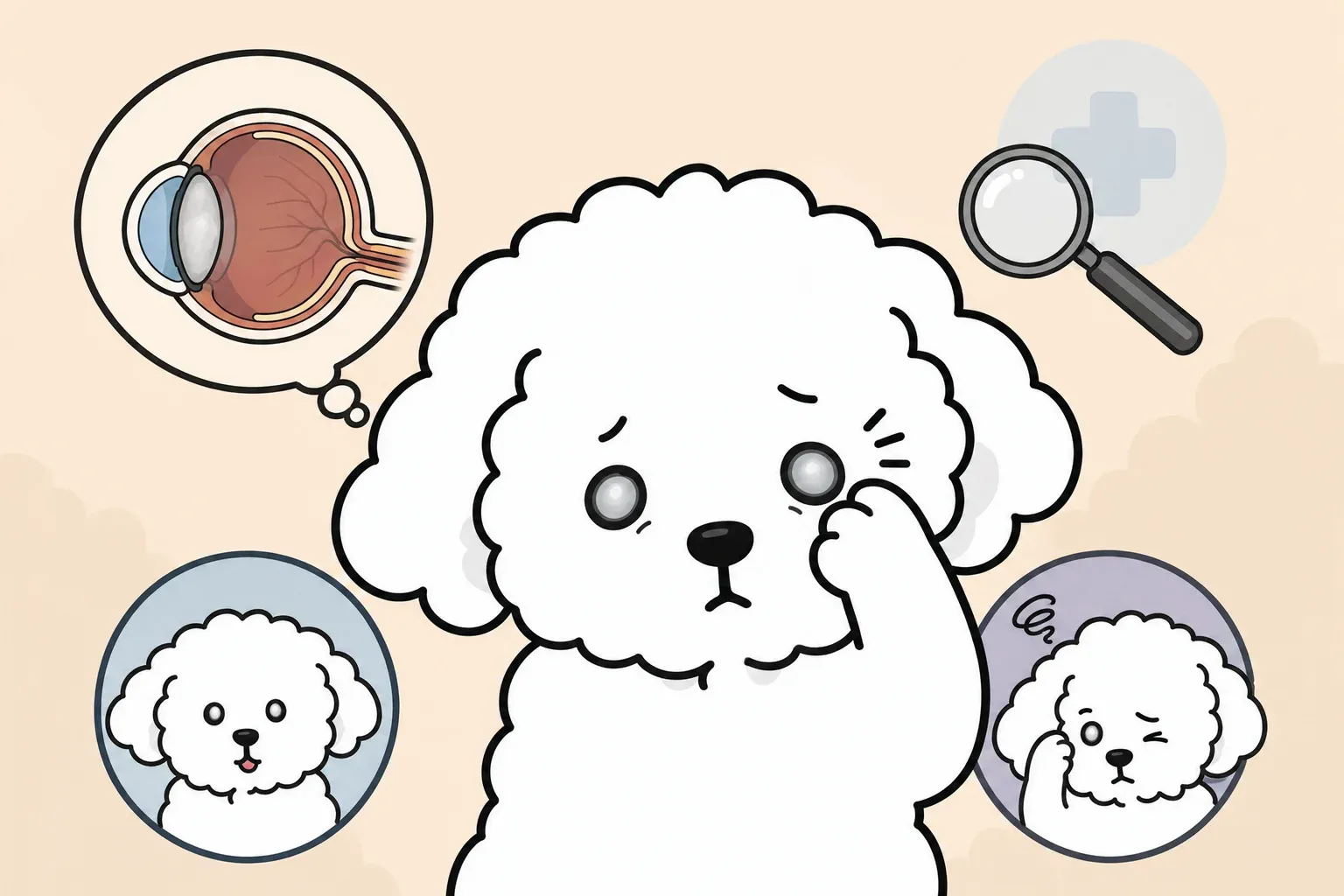
You notice your dog squinting in bright light. Or maybe their eyes have taken on a faint bluish-gray film. You pull out your phone and search “cloudy eyes in dogs”—and immediately land on dozens of pages warning about cataracts and blindness.
Before you spiral, here’s the thing: cloudy eyes in dogs are not always cataracts. One of the most common eye changes in older dogs is an entirely normal aging process that poses no threat to vision. Knowing the difference can save you a great deal of unnecessary worry—and help you catch a real problem when one actually exists.
This guide covers everything dog owners need to understand about dog cataracts symptoms, how they progress, what causes them, and what your options are when a true cataract is confirmed.
Cloudy Eyes Don’t Always Mean Cataracts
When a middle-aged or older dog develops a bluish-gray haze over the eye, the most frequent culprit is not a cataract—it is nuclear sclerosis (also called lenticular sclerosis). Understanding this distinction is the starting point for any conversation about dog eye health.
Nuclear Sclerosis: A Normal Part of Aging
Nuclear sclerosis is a hardening and increased density of the center (nucleus) of the lens that occurs naturally as dogs age. The lens fibers that have been accumulating since birth gradually compress, creating an optical change that appears as a subtle gray or blue haze when light hits the eye at certain angles.
The critical point: nuclear sclerosis does not meaningfully impair vision. Most dogs with nuclear sclerosis navigate their environment normally, respond to hand signals, and pass basic obstacle tests without difficulty. The change is cosmetic from a functional standpoint.
Nuclear sclerosis typically appears in dogs around 6–7 years of age and is nearly universal in dogs over 10. Both eyes are usually affected simultaneously, and the progression is gradual and consistent over years.
Cataracts, by contrast, involve structural opacity within the lens that blocks light from reaching the retina. Depending on how much of the lens is affected, cataracts can range from subtle vision blur to complete blindness.
How to Tell the Difference: The Flash Test
A simple at-home check that can give you a preliminary clue is the tapetal reflection test, sometimes called the flash test.
In a dimly lit room, shine a small flashlight or use your phone’s flashlight into your dog’s eye from about 12 inches away. Observe what happens behind the haze:
- Nuclear sclerosis: The light passes through the haze and you can still see the characteristic greenish-yellow glow (tapetal reflection) from the back of the eye. The lens looks uniform and translucent.
- Cataract: The opacity blocks or fragments the light. The reflection may appear absent, patchy, or irregular. The lens looks distinctly white or opaque rather than simply hazy.
This test is a useful screening tool, not a diagnosis. Veterinary examination with a slit lamp (biomicroscope) is required to differentiate the two conditions definitively and to assess whether vision is affected. If the flash test gives you pause, schedule a veterinary visit—do not rely on the at-home check alone.
What Are Cataracts in Dogs?
A cataract is an opacity (loss of transparency) within the crystalline lens of the eye. Unlike nuclear sclerosis, cataracts represent pathological change—protein structures within the lens break down or aggregate abnormally, scattering light and reducing how much reaches the retina.
How the Lens Works and Why It Clouds
The lens sits directly behind the iris (the colored part of the eye) and pupil. Its primary function is to focus light precisely onto the retina, where photoreceptor cells convert it into neural signals the brain interprets as vision.
The lens is composed almost entirely of specialized proteins called crystallins arranged in a highly ordered, transparent structure. Maintaining this transparency requires a continuous supply of glucose, a delicate balance of electrolytes, and protection from oxidative stress (cellular damage from reactive oxygen molecules).
When any of these conditions are disrupted—through genetic mutation, metabolic disease like diabetes, inflammation, trauma, or accumulated UV damage—the crystallin proteins become disorganized, aggregate into clumps, or undergo chemical changes that make them opaque. The result is a cataract: a lens that scatters rather than focuses light.
The 4 Stages of Canine Cataracts
Veterinary ophthalmologists classify cataracts by the percentage of the lens they occupy, which directly correlates to vision impact:
| Stage | Lens Involvement | Vision Impact |
|---|---|---|
| Incipient | Less than 15% | Minimal to none; detected only with slit lamp |
| Immature | 15–99% (partial) | Reduced visual acuity; some light/shape perception |
| Mature | 100% (complete) | Functional blindness in affected eye |
| Hypermature | Shrinking, liquefying lens | Severe vision loss; high risk of secondary uveitis and glaucoma |
The progression from incipient to mature can take months to years depending on the underlying cause—with one critical exception. Diabetic cataracts can advance from incipient to mature within days to weeks due to the rapid accumulation of sorbitol (a sugar alcohol) inside the lens.
Why hypermature cataracts require urgent attention: As the lens protein liquefies, it leaks through the lens capsule and triggers an immune-mediated inflammatory reaction called lens-induced uveitis (LIU). Chronic uveitis damages the intraocular structures and dramatically increases the risk of glaucoma. Both conditions can cause permanent blindness and significant pain, independent of the cataract itself.
What Causes Cataracts in Dogs?
Unlike humans, where UV exposure and aging are the dominant causes, cataracts in dogs are most frequently hereditary. Understanding the cause matters because it affects prognosis, treatment planning, and decisions about breeding.
Hereditary Cataracts and High-Risk Breeds
Hereditary cataracts (HC) arise from genetic mutations that alter lens protein structure and are the most common cause of cataracts in dogs. The ACVO (American College of Veterinary Ophthalmologists) maintains a Companion Animal Eye Registry (CAER) that documents breed-specific eye disease prevalence.
High-risk breeds include:
- Cocker Spaniel (American and English): One of the highest-prevalence breeds in US veterinary ophthalmology statistics; cataracts can develop as early as 1–2 years of age
- Labrador Retriever: Posterior polar cataracts with a known genetic marker (HSF4 gene mutation) identified in research
- Poodle (Toy, Miniature, Standard): Frequently affected; posterior subcapsular cataracts common
- Boston Terrier: High prevalence of posterior Y-suture cataracts
- Siberian Husky: Hereditary posterior cataracts documented; CAER certification recommended for breeding dogs
- Bichon Frise: Posterior cortical cataracts, often bilateral
- Golden Retriever: Moderate risk; typically develops in older age
Hereditary cataracts are typically bilateral (affecting both eyes), though one eye often progresses faster than the other. They tend to develop in young to middle-aged dogs, distinguishing them from age-related nuclear sclerosis.
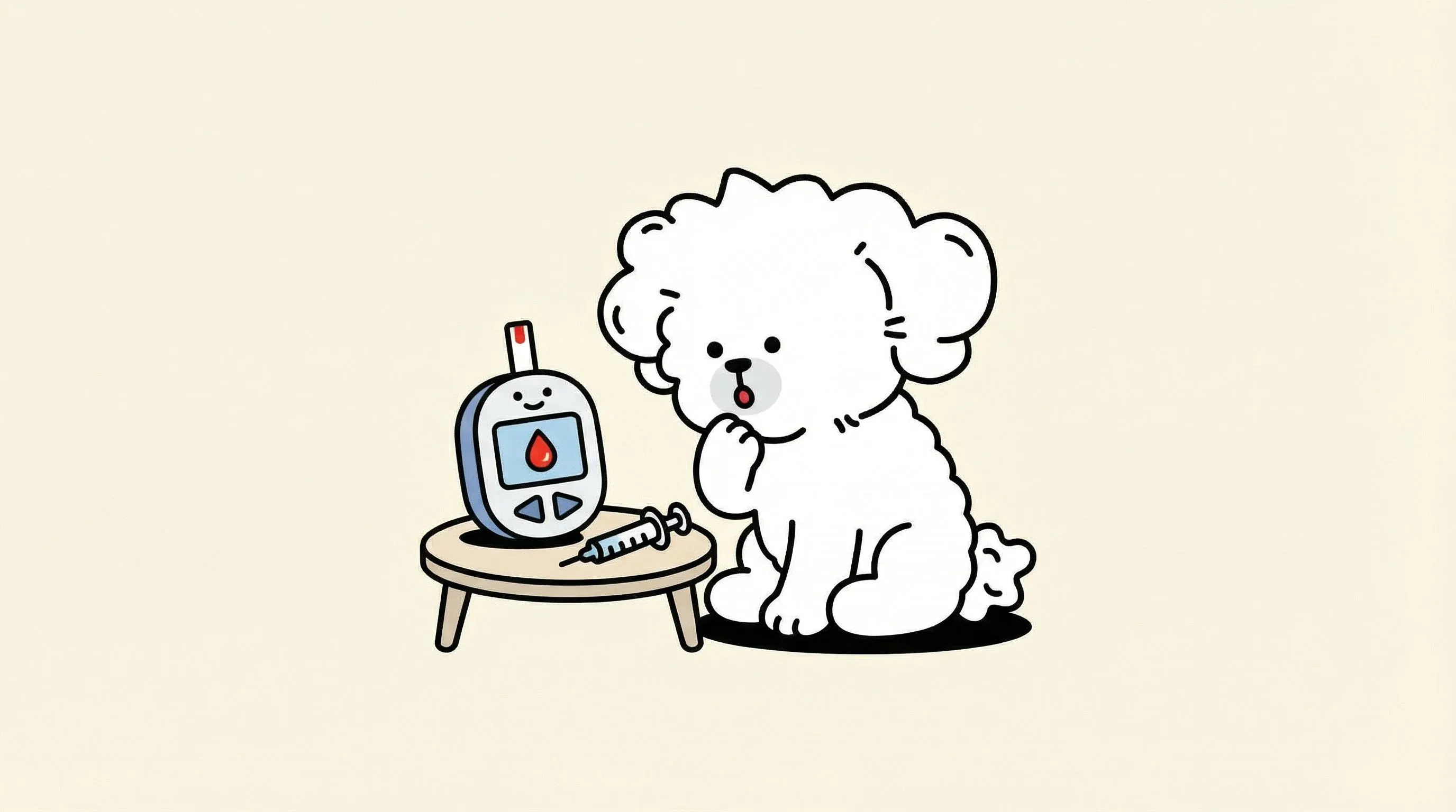
Diabetes-Related Cataracts
Diabetic cataracts represent a medical emergency within the cataract category. Approximately 75% of diabetic dogs will develop cataracts within a year of diabetes diagnosis, and the progression is uniquely rapid.
The mechanism: when blood glucose levels are chronically elevated, excess glucose enters the lens and is converted by an enzyme called aldose reductase into sorbitol, a sugar alcohol the lens cannot excrete efficiently. Sorbitol accumulates, drawing water into the lens via osmosis. This rapid fluid influx disrupts the lens fiber structure—sometimes causing visible opacity within 24–48 hours of significant blood sugar elevation.
For dogs with diagnosed diabetes mellitus, proactive ophthalmologic monitoring is essential, not optional. Glycemic control directly influences cataract development rate; poorly controlled diabetes accelerates progression significantly.
To understand how blood sugar management connects to eye health, see our guide on dog diabetes management.
Age, Trauma, UV Exposure, and Other Causes
Age-related cataracts: Distinct from nuclear sclerosis, senile cataracts do occur in older dogs due to cumulative oxidative damage to lens proteins. These typically develop after age 8–9 and progress slowly.
Traumatic cataracts: Blunt trauma to the eye, penetrating injuries, or electric shock can disrupt the lens capsule or alter lens metabolism, leading to focal or diffuse opacity in the affected eye.
UV exposure: Chronic UV light exposure damages lens proteins through photooxidation. Outdoor dogs with prolonged sun exposure over many years may have accelerated oxidative lens changes, though this is rarely the sole cause in dogs.
Secondary cataracts: Chronic uveitis (intraocular inflammation from infectious diseases like ehrlichiosis, toxoplasmosis, or immune-mediated conditions), retinal degeneration, and certain medications can cause cataracts as secondary complications.
Nutritional deficiencies: Historically, cataracts were documented in puppies fed severely arginine-deficient diets. Modern commercial dog foods formulated to AAFCO standards make nutritional cataracts rare, but they remain a consideration in dogs on homemade diets without proper supplementation.
Recognizing the Early Signs
Cataracts in dogs are frequently more advanced by the time owners notice them than owners realize—which is why behavioral changes often precede the obvious visual finding.
Behavioral Changes to Watch For
The following behavioral changes, particularly in combination, should prompt an eye examination:
- Bumping into objects, especially new or rearranged furniture, or objects in familiar spaces
- Hesitation on stairs, particularly when descending or in dim lighting
- Reluctance to navigate new environments, especially at dusk or in low-light conditions
- Startling more easily when approached from certain angles (typically the affected side)
- Reduced interest in fetching or other vision-dependent activities
- Squinting or increased blinking in bright light
- Increased eye rubbing or pawing at the face (may indicate secondary uveitis)
- Changes in depth perception: misjudging distances, hesitating before jumping
Many owners attribute these changes to aging or arthritis. A simple at-home vision screen—rearranging a few objects in a familiar room and observing navigation—can reveal vision deficits that may not be obvious in the dog’s usual environment.
Symptoms by Stage
The behavioral picture changes with cataract progression:
Incipient stage: No behavioral symptoms. The opacity is too small to meaningfully affect vision. Detected only during routine veterinary eye exam or CAER screening.
Immature stage: Subtle signs emerge—mild hesitation in new environments, slight decrease in activity in dim conditions. Vision is reduced but functional. This is an important window for surgical consultation because the lens is still intact and surgical outcomes are best.
Mature stage: Functional blindness in the affected eye. If both eyes are involved, the dog may walk into objects, become anxious in unfamiliar spaces, and show marked behavioral changes. Many dogs adapt remarkably well to single-eye vision loss.
Hypermature stage: Blindness, often accompanied by signs of uveitis—redness, tearing, squinting, and pain. This stage requires urgent veterinary attention to manage secondary complications regardless of surgical decisions.
Diagnosis: What to Expect at the Vet
A routine physical exam is not sufficient to fully evaluate cataracts. Your veterinarian will perform or refer for several specialized tests.
Slit-Lamp Exam, Tonometry, and Ultrasound
Slit-lamp biomicroscopy is the foundational tool. The slit lamp projects a thin beam of light through the lens, allowing the examining veterinarian to assess the location, density, and extent of the opacity with precision unavailable to the naked eye. This exam definitively distinguishes cataracts from nuclear sclerosis.
Tonometry measures intraocular pressure (IOP). Normal IOP in dogs is approximately 15–25 mmHg. Elevated pressure indicates glaucoma—a secondary complication that can develop quickly with mature or hypermature cataracts and that requires immediate treatment. This measurement is essential before any surgical planning.
Ocular ultrasound (B-scan) becomes necessary when the cataract is dense enough to prevent direct visualization of the retina. The retina must be functionally intact for surgery to restore vision—if the retina has detached or degenerated, restoring lens transparency will not improve sight. Ultrasound can reveal retinal detachment, vitreous disease, or subluxation (displacement) of the lens.
Electroretinogram (ERG) measures electrical activity in the retina in response to light stimulation. This test is specifically used pre-surgically to confirm retinal function in eyes with mature cataracts where direct fundoscopic examination is impossible. A dog with retinal degeneration (such as progressive retinal atrophy) would not benefit from cataract surgery; ERG identifies this before the procedure.
Most general practitioners will perform the initial examination and refer to a board-certified veterinary ophthalmologist (DACVO) for advanced diagnostics and surgical consultation.
At-Home Checks and Their Limitations
Beyond the flash test described earlier, there are a few additional at-home observations that can be informative:
Maze test: Set up a simple obstacle course using cardboard boxes or cushions in a familiar room. First observe your dog navigating in normal lighting, then in dim lighting. Vision-impaired dogs often perform worse in low light because they rely more heavily on spatial memory in their regular environment.
Menace response: While squinting one eye closed, move your hand rapidly toward the open eye from the side. A dog with intact vision will blink or pull back. This tests the visual pathway but is not reliable in very calm or stoic dogs.
Limitations: At-home checks can suggest a problem but cannot replace veterinary diagnosis. They cannot differentiate between cataract types, measure disease severity, detect secondary complications, or assess retinal function.
Treatment Options
Once a cataract diagnosis is confirmed, treatment decisions are guided by the stage of disease, the dog’s overall health, and practical factors including the owner’s capacity for post-operative care.
Phacoemulsification Surgery: Procedure and Success Rates
Phacoemulsification (phaco) is the veterinary equivalent of the cataract surgery performed in humans—and the results are similarly successful. The procedure involves:
- Small incisions in the cornea
- Insertion of a probe that uses ultrasonic vibrations to emulsify (break apart) the opaque lens material
- Removal of the emulsified material by suction
- Implantation of an artificial intraocular lens (IOL) into the preserved lens capsule
- Closure of incisions with absorbable sutures
The procedure is performed under general anesthesia by a board-certified veterinary ophthalmologist and typically takes 45–90 minutes per eye.
Success rates: Large case series published in the Journal of Veterinary Ophthalmology report 90–95% of dogs having vision in the surgical eye at the 12-month follow-up. The best outcomes occur when:
- Surgery is performed during the immature stage (before the lens becomes hypermature)
- The retina is confirmed functional by pre-operative ERG
- Uveitis is well-controlled before surgery
- Post-operative drop compliance is excellent
Outcomes are significantly poorer when surgery is delayed until the hypermature stage due to increased surgical complexity and irreversible secondary damage.
Surgery Costs and Realistic Considerations
Cataract surgery in dogs in the United States typically ranges from $2,800 to $4,900 per eye, including pre-operative diagnostics (ERG, ultrasound, blood work), the surgical procedure, and immediate post-operative care. Most veterinary ophthalmology practices charge separately for ongoing follow-up visits.
This represents a substantial financial commitment. When evaluating the decision, consider:
For the surgery:
- Phaco surgery is the only proven method to restore functional vision once cataracts are mature
- Prevention of painful secondary complications (uveitis, glaucoma) that can develop without intervention
- Studies show dogs maintain good quality of life and owners report high satisfaction with surgical outcomes
Against the surgery (or requiring further evaluation):
- Dogs with poor anesthetic risk due to cardiac or respiratory disease
- Active, uncontrolled uveitis (surgery in an inflamed eye has higher complication rates)
- Retinal degeneration identified by ERG (surgery will not restore vision if the retina cannot process light)
- Uncontrolled diabetes (glycemic stabilization is required before surgical clearance)
- Owner inability to maintain the intensive post-operative eye drop schedule (4–6 times daily for several weeks)
The “is it worth it for a senior dog?” question: Age alone is not a contraindication. A healthy 13-year-old dog with well-controlled health conditions and good anesthetic risk may be an excellent surgical candidate. The evaluating question is not age but physiologic health status. Discuss anesthetic risk assessment explicitly with your veterinarian.
Surgical consultation with a DACVO specialist typically costs $150–$350 and provides the clearest picture of whether your dog is a good candidate before you make a financial commitment.
Non-Surgical Management: Eye Drops, Nutrition, and Environment
For dogs who are not surgical candidates, whose cataracts are in early stages, or whose owners choose conservative management, there are meaningful steps to slow progression and protect quality of life.
Anti-inflammatory management: The most medically important non-surgical intervention is controlling secondary uveitis. Veterinarians commonly prescribe topical corticosteroids or NSAIDs (such as prednisolone acetate or diclofenac ophthalmic drops) to reduce lens-induced intraocular inflammation. This does not treat the cataract itself but prevents or delays the destructive secondary complications.
Investigational drops: N-acetylcarnosine (NAC) eye drops and lanosterol drops have received attention as potential non-surgical treatments. Peer-reviewed evidence in dogs remains limited and inconsistent—neither is approved by the FDA for this use in veterinary medicine. If your veterinarian mentions these as adjuncts, ask about the current evidence base.
Environmental adaptation for visually impaired dogs: Dogs with reduced vision adapt remarkably well when their environment is consistent:
- Keep furniture arrangement stable
- Use baby gates to block stairs
- Place textured mats to signal zone transitions (rugs at doorways, food bowl locations)
- Use scent markers (a drop of vanilla extract near the water bowl, for example) to help orientation
- Speak before approaching to avoid startling
- Keep outdoor spaces well-defined with fencing
Nutritional support: Antioxidant nutrients protect against oxidative lens damage and may help slow progression in early-stage disease. These are discussed in the Prevention section below.
Post-Surgery Recovery and Home Care
Phacoemulsification requires a committed recovery period. Owners who understand the protocol before surgery report better compliance and outcomes.
Recovery Timeline and Restrictions
Days 1–7 (Acute phase):
- The dog must wear an Elizabethan collar (e-collar) continuously—including during sleep. This is non-negotiable; any rubbing or pawing can displace the IOL or open incisions.
- Eye drops are typically administered 4–6 times daily. Drops usually include a topical antibiotic, a corticosteroid or NSAID, and a mydriatic (pupil-dilating agent). Missing doses significantly increases complication risk.
- No running, jumping, rough play, or swimming. Leash-only bathroom breaks.
- The first recheck is typically 24–48 hours post-surgery.
Weeks 2–4 (Subacute phase):
- Drop frequency gradually tapers (usually from 4–6 times to 3–4 times daily).
- Activity restrictions begin to ease but remain significant—no swimming, no off-leash running.
- Recheck appointments at week 2 and week 4.
Weeks 5–8:
- Most dogs are down to 2–3 drops per day.
- Normal activity usually resumes by week 6–8 if recovery is uneventful.
- Recheck at 6–8 weeks.
Long-term:
- Some dogs require lifelong topical anti-inflammatory therapy to prevent chronic uveitis.
- Annual recheck by veterinary ophthalmologist is recommended.
The most common owner challenges are e-collar compliance (dogs find them distressing) and drop schedule maintenance. Planning a support system in advance—especially if you work outside the home—is important.
Preventing Recurrence
Posterior capsule opacification (PCO)—clouding of the lens capsule that remains after phaco—occurs in some dogs and may require a brief laser procedure (Nd:YAG laser capsulotomy) to clear vision again. This is performed without anesthesia in most cases.
Cataracts in the remaining healthy lens material (anterior capsule) are uncommon when surgery is performed before the lens becomes hypermature.
The lens that was removed cannot regrow—but new cataracts cannot technically form in a successfully implanted IOL. The recurrence concern centers on secondary opacification and ongoing management of uveitis.
Prevention and Daily Eye Care
While hereditary cataracts cannot be prevented (genetic testing and responsible breeding practices reduce their frequency in future generations), there are practical steps that can delay progression, reduce oxidative stress, and support overall eye health.
Eye-Healthy Nutrients
The lens is particularly vulnerable to oxidative damage because it has limited ability to regenerate damaged proteins. Antioxidant nutrients help neutralize reactive oxygen species before they damage lens structures.
Key antioxidants for lens protection:
- Lutein and zeaxanthin: Carotenoids that accumulate in ocular tissue and filter high-energy blue light. Found in leafy greens; available as supplements.
- Astaxanthin: A potent carotenoid antioxidant with strong evidence for tissue protection; crosses the blood-retinal barrier.
- Vitamin E (tocopherols): Fat-soluble antioxidant; protects lens cell membranes from lipid peroxidation.
- Vitamin C (ascorbate): Found in high concentrations in the aqueous humor (fluid surrounding the lens); supports lens protein stability.
- Beta-carotene: Converted to Vitamin A; essential for retinal photoreceptor function.
- Omega-3 fatty acids (EPA/DHA): Reduce systemic and ocular inflammation; DHA is a structural component of retinal photoreceptors.
For breed-specific supplement recommendations and supporting eye health through diet, see our guide on dog eye health supplements.
Connecting eye nutrition to overall senior health is also valuable—senior dog diet and nutrition covers how nutrient needs shift as dogs age.
UV Protection and Eye Hygiene
UV protection: Dogs with outdoor lifestyles—particularly those in high-altitude or high-UV environments—accumulate photooxidative stress over time. Canine sunglasses (doggles) offer UV protection and are well-tolerated by many dogs when introduced gradually. Limiting peak-sun exposure (10 a.m.–3 p.m.) during summer months is a practical alternative. For a full overview of breed-specific UV risks and safe sunscreen selection, see our dog sunburn and UV protection guide.
Eye hygiene: Chronic discharge or tear staining around the eye can harbor bacterial growth and increase the risk of secondary infections that cause uveitis. Gently wiping the periocular area with a damp cloth or veterinarian-approved eye wipe daily is appropriate for dogs prone to discharge.
For comparison and context around other canine eye conditions, our guide on dog cherry eye treatment explains another common eye condition that requires veterinary intervention.
Diabetes management: For diabetic dogs, rigorous glycemic control is the single most powerful cataract prevention strategy. Every period of elevated blood glucose accelerates sorbitol accumulation in the lens. Working closely with your veterinarian on insulin dosing, diet, and monitoring is as important for your diabetic dog’s eyes as it is for their systemic health.
Annual eye screening: For high-risk breeds, annual CAER examinations by a board-certified ophthalmologist from 1–2 years of age allow detection at the incipient stage, when surgical outcomes are optimal and preventive strategies can be maximized. Cataracts are just one of many conditions affecting dog eyes — for a broader overview of symptoms, urgency levels, and other common eye diseases, see our dog eye diseases symptoms guide.
Age-related changes and cognitive health: Senior dogs experiencing vision changes may show behavioral shifts that overlap with cognitive dysfunction. Understanding the difference helps owners respond appropriately—see our guide on dog cognitive dysfunction for more.
References
- 1. Cataracts in Dogs - Veterinary Ophthalmology (Gelatt)
- 2. American College of Veterinary Ophthalmologists (ACVO) - Breed-Specific Eye Disease Registry
- 3. Cataracts in Dogs - Merck Veterinary Manual
- 4. Diabetic Cataracts in Dogs - Veterinary Clinics of North America
- 5. Phacoemulsification outcomes in dogs - Journal of Veterinary Ophthalmology
FAQ
What happens if cataracts in dogs are left untreated?
Is cataract surgery worth it for a senior dog?
Can cataracts in dogs be treated with eye drops alone?
Do cataracts always affect both eyes in dogs?
What foods support eye health in dogs?
At what age should cataract screening begin for high-risk breeds?
Related Articles

Dog Eye Supplement Guide: What Veterinary Research Says
Which dog eye supplement ingredients are research-backed? Lutein, astaxanthin, and omega-3 explained with dosage guidance and quality verification tips.
Is Your Dog Drinking Too Much Water? Diabetes Signs and Blood Sugar Management Guide
Dog diabetes symptoms, insulin therapy, diet management, home blood sugar monitoring, and complication prevention — a vet-backed owner's guide.

Cherry Eye in Dogs: Causes, Treatment, and Surgery Costs
Cherry eye in dogs won't resolve on its own. Learn causes, at-risk breeds, surgical options, realistic US costs ($300–$2,500), and recovery what to expect.