Dog Eye Diseases: 8 Common Conditions Every Owner Should Recognize
Your dog woke up with a swollen, weeping eye — and you’re not sure if you should rush to the emergency vet or wait until morning. It’s one of the most anxiety-inducing moments for dog owners, because the eye is both fragile and non-negotiable: delayed treatment for the wrong condition can mean permanent vision loss.
This guide covers the 8 most common dog eye diseases symptoms, explains how to read their urgency, and gives you a practical framework for what to do — and what not to do — before you get professional help.
Understanding Your Dog’s Eye Anatomy
Before diving into diseases, a brief orientation helps you communicate more precisely with your vet and understand why certain breeds are more vulnerable.
Key Structures: Cornea, Conjunctiva, Lens, and Retina
The cornea is the clear, dome-shaped surface covering the front of the eye. Because it has no blood supply, it depends entirely on tear film for oxygen and nutrients — which is why disruptions to tear production cause so much damage so quickly.
The conjunctiva is the pink mucous membrane lining the inner eyelids and the white of the eye (sclera). It’s the most common site of infection and inflammation. The third eyelid (nictitating membrane), visible in the inner corner, acts as an additional barrier and produces up to 30% of tear film — which is why cherry eye (prolapse of the third eyelid gland) affects tear production so dramatically.
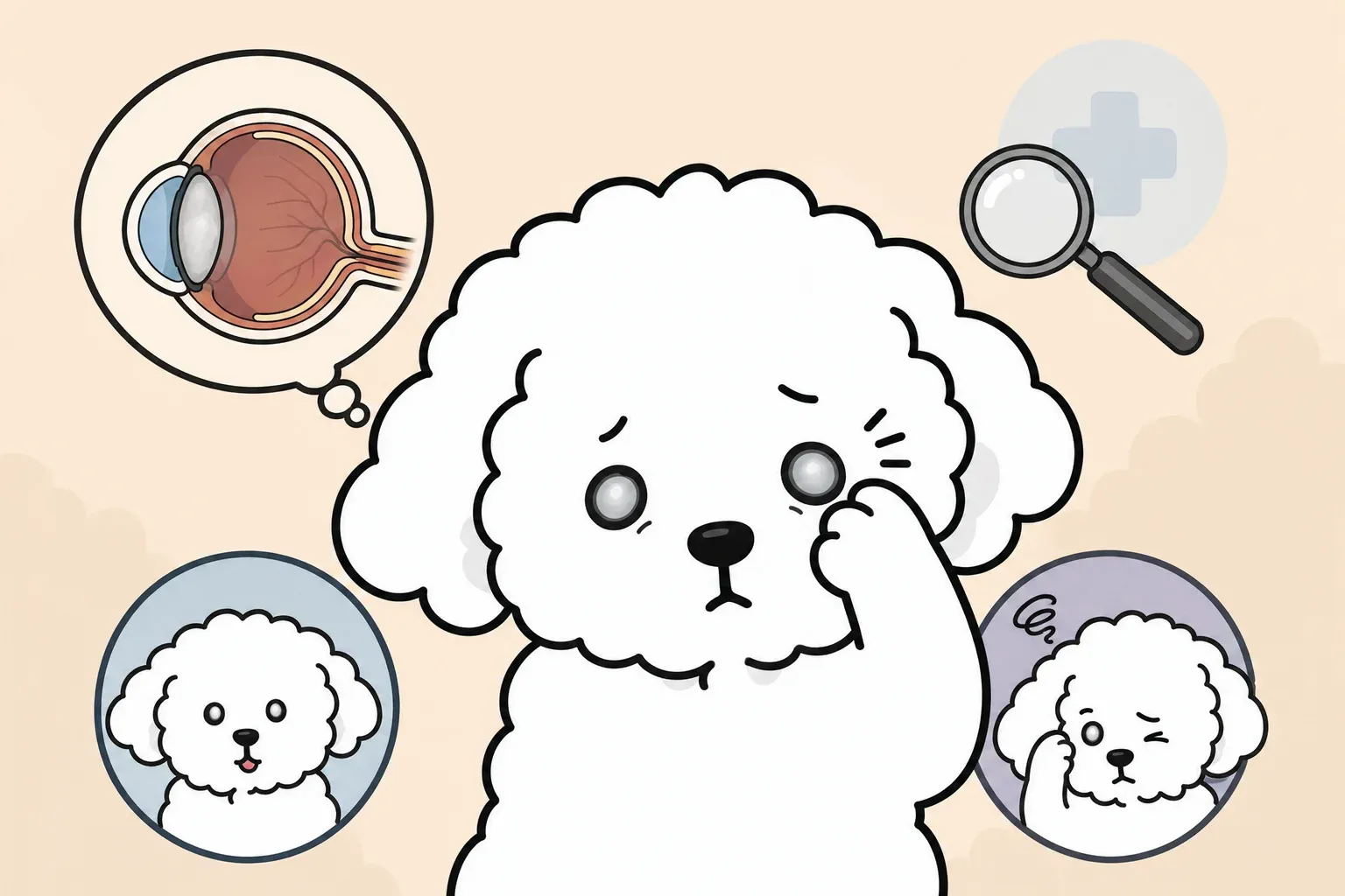
The lens sits behind the iris and focuses light onto the retina, which converts light into signals the brain interprets as vision. Cloudiness in the lens indicates cataracts; cloudiness at the corneal surface points toward different diseases entirely.
Why Dogs Are Prone to Eye Problems
Dogs are closer to the ground than humans, exposing their eyes to grass seeds, dust, and debris during every walk. Facial anatomy plays an even larger role: breeds with flat faces have shallower orbits (eye sockets), so their eyes protrude more and have less natural protection. Genetics add another layer — specific breeds carry inherited mutations affecting intraocular pressure regulation, tear gland function, and retinal health.
7 Warning Signs of Eye Problems in Dogs
Recognizing these signs early is the difference between a simple antibiotic prescription and emergency surgery. Use the table below as a quick reference, then read on for detail on each condition.
| Symptom | Possible Conditions | Urgency |
|---|---|---|
| Red, bloodshot sclera | Conjunctivitis, glaucoma, uveitis | 24 hours (glaucoma: immediate) |
| Yellow or green discharge | Bacterial conjunctivitis, corneal ulcer | 24 hours |
| Clear, watery discharge | Allergic conjunctivitis, epiphora | Monitor |
| Squinting or kept shut | Corneal ulcer, acute glaucoma, uveitis | Immediate–24 hours |
| Cloudiness (cornea) | Corneal ulcer, endothelial degeneration | 24 hours |
| Cloudiness (lens) | Cataracts, nuclear sclerosis | Schedule routine exam |
| Swollen eyelid or tissue | Blepharitis, allergic reaction, abscess | 24 hours |
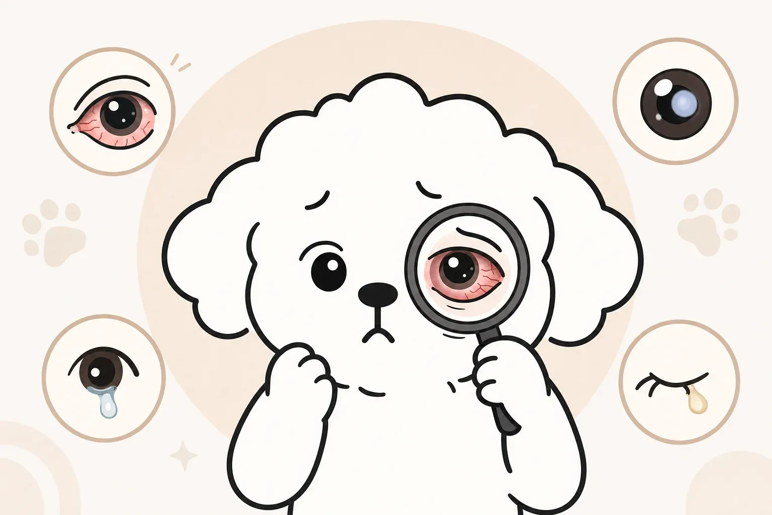
Redness, Discharge, and Excessive Tearing
Redness alone is non-specific — it appears in almost every eye condition. The character of any discharge narrows the differential diagnosis considerably. Clear, watery overflow is most often allergic or anatomical (a blocked nasolacrimal duct). Mucoid (whitish, stringy) discharge is classic for dry eye (KCS). Purulent (yellow-green, thick) discharge points toward bacterial infection or a corneal ulcer with secondary infection.
Excessive tearing that runs down the face and stains the fur is called epiphora. In light-coated breeds, the reddish-brown discoloration comes from porphyrins — iron-containing compounds in tears that oxidize on contact with air. Epiphora itself is a symptom, not a diagnosis; the underlying cause ranges from allergies and blocked tear ducts to entropion (inward-rolling eyelid).
Cloudiness, Squinting, and Swelling
Squinting (blepharospasm) is a pain response. If your dog cannot fully open one eye or keeps rubbing it, assume pain is present until proven otherwise — this is a 24-hour-or-sooner veterinary situation. Cloudiness requires localizing: is it on the surface (cornea) or deep inside (lens)? Surface cloudiness in a painful, red eye almost always means a corneal ulcer or advanced dry eye. Painless, slow-onset cloudiness in a senior dog’s lens is more likely cataracts or nuclear sclerosis.
8 Common Dog Eye Diseases: Causes and Symptoms
Conjunctivitis: The Most Common Eye Infection
Conjunctivitis — inflammation of the conjunctiva — is the most frequent reason dogs visit veterinary ophthalmologists. It rarely threatens vision but causes significant discomfort.
Causes: Bacterial infection (Staphylococcus, Streptococcus), viral infection (canine distemper, herpesvirus), allergies (environmental pollens, dust mites, mold), or mechanical irritation from entropion or foreign material.
Symptoms: Redness of the pink tissue around the eye, discharge ranging from clear to purulent, mild squinting, and sometimes swelling of the conjunctival tissue itself (chemosis). Both eyes affected simultaneously suggests an allergic or systemic cause; one eye only is more consistent with infection or local irritation.
Progression: Bacterial conjunctivitis responds well to topical antibiotics within 5–7 days. Untreated, the infection can extend to the cornea or spread to the other eye.
If your dog has known seasonal allergies, managing environmental allergens reduces the frequency of allergic conjunctivitis significantly.
Corneal Ulcers: Surface Injuries That Need Urgent Care
A corneal ulcer is a defect in the cornea’s surface epithelium — essentially an open wound on the eye. In dogs, they can progress from superficial to full-thickness perforation within 24–48 hours if infected with certain bacteria (Pseudomonas aeruginosa is notorious for rapid tissue destruction).
Causes: Trauma (scratch from a cat, twig, or grass seed), entropion (eyelid rolling inward so lashes rub the cornea), dry eye reducing the protective tear film, foreign bodies trapped under the eyelid, or in Boxers — a breed-specific condition called indolent (Boxer) ulcer that fails to heal normally.
Symptoms: Sudden, intense squinting; excessive tearing; cloudiness or a visible white/gray spot on the cornea; pawing at the eye; reluctance to open the eye in bright light (photophobia). Brachycephalic breeds with prominent eyes are at highest risk of traumatic ulcers.
Progression: Superficial ulcers treated with antibiotic drops and an E-collar typically heal in 5–7 days. Deep or infected ulcers can perforate the eye — a vision-ending emergency.
Dry Eye (KCS): When Tear Production Fails
Keratoconjunctivitis sicca (KCS) — colloquially “dry eye” — occurs when the lacrimal (tear) glands fail to produce adequate watery tears. The tear film normally protects, lubricates, and nourishes the cornea; without it, the surface deteriorates progressively.
Causes: Immune-mediated destruction of lacrimal gland tissue (the most common cause in dogs, distinct from human dry eye), side effects of certain sulfonamide antibiotics, hypothyroidism, distemper, or surgical removal of a cherry eye gland.
Symptoms: Thick, ropy, gray-green discharge (the body overproduces mucin to compensate for the lack of water); dull, lusterless corneal surface; chronic redness; recurrent corneal ulcers; and in advanced cases, brown pigment deposited on the cornea (pigmentary keratitis) that permanently impairs vision.
Affected breeds: Cocker Spaniels, Bulldogs, Pugs, West Highland White Terriers, and Lhasa Apsos have particularly high incidence rates. Learn more about preventive eye nutrition that may support lacrimal gland function.
Treatment: Lifelong cyclosporine or tacrolimus eye drops stimulate lacrimal gland function in immune-mediated cases. Artificial tear supplementation provides symptomatic relief. This condition is manageable, not curable — owners must commit to daily medication for the dog’s life.
Glaucoma: The Silent Vision Thief
Glaucoma results from elevated intraocular pressure (IOP) that damages the optic nerve and retina. It is one of the few eye conditions where hours genuinely matter: acute glaucoma can cause irreversible blindness within 24–72 hours.
Causes: Primary glaucoma (inherited predisposition causing abnormal drainage angle anatomy) or secondary glaucoma (elevated pressure caused by uveitis, lens luxation, tumors, or trauma).
Symptoms of acute attack: Sudden, severe redness; a visibly enlarged eyeball (the eye looks “bigger” than normal); rock-hard feeling when gently pressing the closed eyelid; extreme pain indicated by head pressing, lethargy, or loss of appetite; fixed, dilated pupil; and corneal cloudiness from fluid accumulation.
Predisposed breeds: Cocker Spaniels, Basset Hounds, Chow Chows, Shar-Peis, Siberian Huskies, and Norwegian Elkhounds carry the highest genetic risk for primary glaucoma. Regular IOP screening — measuring the eye’s internal pressure with a tonometer — is recommended annually for these breeds starting at age 3–4.
If your dog seems reluctant to move, is pressing their head against walls, or shows other signs of discomfort, these can be pain behavior indicators worth discussing with your vet promptly.
Blepharitis: Eyelid Inflammation
Blepharitis is inflammation of the eyelid margins — the skin, muscle, and glands that form the lid edge. It’s often underdiagnosed because owners attribute the symptoms to simple conjunctivitis.
Causes: Bacterial infection (typically Staphylococcus), Demodex or Sarcoptes mite infestation, allergic reaction, autoimmune conditions (pemphigus foliaceus), or sebaceous adenitis affecting the meibomian glands (which produce the oily component of tears).
Symptoms: Crusty, thickened eyelid margins; loss of eyelashes or abnormal lash growth; redness and swelling of the lid; secondary conjunctivitis from debris falling onto the eye; and sometimes ulceration of the lid margin in severe cases.
Treatment: Warm compresses to loosen crusts, gentle cleaning of lid margins, antibiotics or antifungals based on culture, and addressing the underlying cause (mite treatment, immunosuppression for autoimmune forms).
Epiphora: Excessive Tear Overflow
Epiphora describes tear overflow onto the face — the visible tear staining seen in many small, light-colored breeds. It’s a symptom of underlying lacrimal system dysfunction rather than a disease itself.
Causes: Anatomical blockage of the nasolacrimal duct (the channel that normally drains tears into the nose), distichia (extra eyelashes growing from abnormal locations), entropion, shallow nasolacrimal puncta (the drainage openings), or any condition that increases tear production (allergies, irritation, early KCS paradoxically in some cases).
Symptoms: Persistent wetness below the eyes; reddish-brown staining on white or light-colored fur; skin irritation or secondary yeast/bacterial infection in the moist skin folds beneath the eyes.
For a detailed management protocol, see dog tear stain care — including cleaning techniques and when staining warrants further veterinary investigation.
Uveitis: Inflammation Inside the Eye
Uveitis — inflammation of the uvea (iris, ciliary body, and choroid) — is significant because it’s frequently a sign of systemic disease. It’s also one of the leading causes of secondary glaucoma and cataracts.
Causes: Infectious diseases (Lyme disease, Rocky Mountain spotted fever, blastomycosis, leptospirosis), immune-mediated conditions, trauma (traumatic uveitis), lens-induced uveitis (from a hypermature cataract leaking proteins), or neoplasia (intraocular tumors).
Symptoms: Redness; a small, constricted (miotic) pupil rather than the dilated pupil of glaucoma; a “cloudy” appearance to the front of the eye (aqueous flare, caused by proteins leaking into the normally clear fluid); squinting; and in chronic cases, color changes to the iris.
Why it matters beyond the eye: Because uveitis so commonly accompanies systemic infection or immune disease, a dog diagnosed with uveitis typically undergoes bloodwork, tick-borne disease panels, and sometimes chest radiographs to identify the underlying cause.
Corneal Endothelial Degeneration: Cloudiness Mistaken for Cataracts
Corneal endothelial degeneration affects the innermost cell layer of the cornea, which pumps fluid out of the corneal tissue to keep it transparent. When these cells die, the cornea swells and turns blue-white — a condition owners frequently mistake for cataracts.
Causes: Age-related cell loss, inherited in certain breeds, or secondary to chronic uveitis or glaucoma.
Symptoms: Progressive, painless blue-white haziness starting at the corneal periphery and moving centrally. The haze is at the surface of the eye (unlike cataracts, which are visible inside the pupil). In advanced cases, the swollen cornea forms fluid-filled blisters (bullous keratopathy) that can rupture and cause corneal ulcers.
Predisposed breeds: Boston Terriers and Chihuahuas have particularly high rates of breed-specific endothelial dystrophy, which often appears in young to middle-aged dogs. Learn more about the distinction between surface and lens cloudiness in the dog cataracts care guide.
Urgency Guide: When to See a Vet
This is the section most competitors don’t provide — a clear triage framework so you’re not guessing at 11 PM whether your dog needs emergency care or can wait until your regular vet opens Monday.
Emergency (Immediate): Acute Glaucoma, Deep Corneal Ulcers, Eye Proptosis
These conditions can cause irreversible blindness within hours. Go to an emergency veterinary clinic now, even if it means leaving in the middle of the night.
Acute glaucoma:
- Sudden-onset, intensely red eye
- Eyeball visibly larger than normal
- Extreme pain (not eating, head pressing, lethargy)
- Fixed, dilated pupil
Deep or perforated corneal ulcer:
- Dog cannot open the eye at all
- Visible dark protrusion at ulcer center (iris prolapse — the inside of the eye pushing through)
- Eye suddenly turned soft or sunken
- Visible blood inside the eye (hyphema)
Eye proptosis (eyeball displaced from socket):
- Eyeball visibly out of socket — common trauma result in brachycephalic breeds
- Emergency: keep the eye moist with sterile saline or plain water and transport immediately
Urgent (Within 24 Hours): Persistent Redness, Heavy Discharge, Inability to Open Eye
These situations are not immediately blinding but can deteriorate quickly without treatment. Call your regular vet first thing in the morning; if closed, use an urgent care veterinary clinic.
- Moderate-to-heavy yellow or green discharge
- Eye held partially closed for more than a few hours
- Squinting that doesn’t resolve after gentle cleaning
- Visible white or gray spot on the cornea
- Redness that persists more than 12 hours
- A foreign body you can see but cannot safely remove
Monitor at Home: Mild Tear Staining, Temporary Redness After Play
Some situations can safely be observed for 24–48 hours with home care before scheduling a routine appointment.
- Light, clear discharge that clears with gentle wiping
- Brief redness (less than 2 hours) after rough play or swimming, with no squinting
- Mild seasonal eye irritation in a dog with known allergies, with no change in discharge character
- Established tear staining (reddish-brown fur under the eye) with no new symptoms
Home monitoring rule: If the situation is not clearly improving within 24–48 hours, or if any single “Emergency” or “Urgent” symptom appears, escalate to veterinary care.
Breed Predispositions to Eye Diseases
Understanding your dog’s inherited risks allows you to recognize problems earlier and arrange appropriate preventive screening.
Brachycephalic Breeds (Bulldogs, Pugs, French Bulldogs, Shih Tzus, Boston Terriers): Corneal Ulcers, Proptosis
Flat-faced breeds have shallow orbital rims that leave more of the eye exposed. The combination of prominent eyes, wide palpebral fissures (eyelid openings), and often poor eyelid closure creates a constellation of risks:
- Corneal ulcers: Highest incidence of any breed group. Their exposed corneas are scratched by grass, dust, and even by the skin folds around their nose (nasal fold trichiasis).
- Proptosis: Any trauma — even being grabbed by the scruff — can displace the eyeball from the socket. Know the nearest 24-hour emergency vet clinic before you ever need it.
- Dry eye (KCS): Bulldogs and Pugs have high rates of immune-mediated KCS.
- Entropion: Particularly in Shih Tzus and Bulldogs, the lower eyelid may roll inward.
Small Breeds (Maltese, Poodles, Cocker Spaniels, Lhasa Apsos): Epiphora, Dry Eye, Cataracts
Small breeds account for a disproportionate share of tear-staining complaints, but the underlying causes vary:
| Breed | Primary Eye Risks |
|---|---|
| Maltese | Epiphora (tear staining), blocked nasolacrimal duct |
| Toy/Miniature Poodle | Early-onset hereditary cataracts, progressive retinal atrophy (PRA) |
| Cocker Spaniel | Glaucoma, KCS, cherry eye, distichia |
| Lhasa Apso | KCS, distichia, entropion |
| Shih Tzu | Corneal ulcers, distichia, entropion, KCS |
Annual eye exams are particularly valuable for Cocker Spaniels given their combined glaucoma and KCS risk — both conditions can exist simultaneously, complicating treatment (glaucoma medications and KCS treatments sometimes work against each other).
Large Breeds and Senior Dogs: Glaucoma, Cataracts, Retinal Diseases
Larger breeds and mixed-breed dogs over age 7 face increasing risk from age-related and hereditary conditions:
- Golden Retrievers: Hereditary cataracts (can appear as early as age 2), pigmentary uveitis (a breed-specific inflammatory syndrome), and progressive retinal atrophy.
- Labrador Retrievers: Progressive retinal atrophy (PRA), hereditary cataracts.
- Siberian Huskies: Hereditary cataracts, corneal dystrophy, uveodermatological syndrome.
- German Shepherds: Chronic superficial keratitis (pannus) — an immune-mediated condition causing progressive corneal pigmentation and scarring that responds to topical cyclosporine.
For all dogs over age 7, an annual ophthalmic exam that includes IOP measurement catches glaucoma and early cataract progression before symptoms become obvious. For breed-specific cataract management and staging, the dog cataracts care guide covers surgical candidacy assessment in detail.
Dog Eye Care at Home: Do’s and Don’ts
Home care does not replace veterinary treatment, but it can reduce irritation, remove infectious material, and prevent self-trauma between vet visits. Done incorrectly, it can worsen conditions dramatically.
Safe Eye Cleaning Techniques
What you need:
- Sterile, preservative-free saline solution (contact lens saline is appropriate)
- Soft, non-fibrous pads (cotton balls shed fibers; sterile gauze or dedicated eye wipes are better)
- E-collar (cone) if your dog is pawing at the eye
Step-by-step cleaning:
- Wash your hands thoroughly.
- Gently hold your dog in your lap or have a helper steady them — force makes eye cleaning dangerous.
- Moisten the pad with saline. Do not pour directly into the eye unless explicitly instructed by your vet.
- Starting from the inner corner (nearest the nose), wipe outward in a single motion. Never wipe inward toward the nose, which can introduce bacteria.
- Use a fresh pad for each wipe — reusing spreads discharge back onto the eye.
- Clean the surrounding fur, especially the skin fold below the eye in brachycephalic breeds, to prevent secondary skin infection from moist debris.
Frequency: Once or twice daily for dogs with ongoing discharge. More frequent cleaning doesn’t speed healing and increases irritation risk.
Grooming and Environmental Management
- Trim facial fur that touches or hangs over the eyes — particularly in Shih Tzus, Lhasa Apsos, and Poodles. Long hair wicks moisture onto the cornea and acts as a mechanical irritant. Use blunt-tipped scissors and have a helper; never groom the eye area alone.
- Avoid walking in high pollen environments during allergy season for dogs with a history of allergic conjunctivitis.
- Swimming precautions: Rinse eyes with plain saline after swimming in pools (chlorine) or natural bodies of water (bacteria, algae). Dogs with brachycephalic anatomy should wear protective goggles (properly fitted, not excessively tight) during water activities.
- Dust and smoke: Both are common conjunctival irritants. Avoid exposing dogs to cigarette smoke entirely — it’s a proven risk factor for multiple respiratory and eye conditions.
Common Mistakes to Avoid
Do not use human eye drops unless specifically prescribed by your veterinarian. This includes:
- Visine and similar decongestant drops (vasoconstrictors that mask symptoms)
- OTC antihistamine eye drops (formulations differ from veterinary versions)
- Antibiotic drops prescribed for humans (dosing and drug choice differ)
- Eye drops containing steroids without confirmed diagnosis — steroids can cause catastrophic progression of undetected corneal ulcers
Do not attempt to remove foreign bodies embedded in the cornea or beneath the eyelid without veterinary guidance. Flushing with saline is safe; probing with fingers, cotton swabs, or tweezers is not.
Do not delay the E-collar. If your dog is pawing at or rubbing the eye, put the cone on immediately. A few minutes of self-trauma can turn a superficial ulcer into a perforation.
Do not assume improvement equals resolution. A dog that seems more comfortable after a day of home cleaning may have simply adapted to the discomfort — the underlying pathology continues. Scheduled veterinary follow-up remains necessary.
Nutrition plays a long-term role in maintaining ocular surface health. For research-backed information on antioxidants and omega-3 supplementation, consult the dog eye health supplement guide.
References
FAQ
What does it mean when my dog's eye is red and has discharge?
Can I use human eye drops on my dog?
How do I tell if my dog's cloudy eye is cataracts or something else?
Which dog breeds are most prone to eye problems?
Is dog eye disease an emergency?
Related Articles
Cloudy Eyes in Dogs: Is It Always Cataracts?
Dog cataracts symptoms, stages, causes, and treatment options explained. Learn to tell cataracts from nuclear sclerosis—and when surgery is worth it.

Cherry Eye in Dogs: Causes, Treatment, and Surgery Costs
Cherry eye in dogs won't resolve on its own. Learn causes, at-risk breeds, surgical options, realistic US costs ($300–$2,500), and recovery what to expect.
3 Health Warning Signs Hidden in Your Dog's Tear Stains
Dog tear stains signal more than cosmetic issues. Learn to identify root causes—nasolacrimal obstruction, allergies, diet, infection—and apply cause-specific management strategies.